
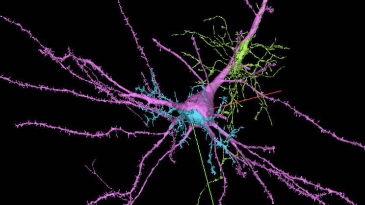
Mapping of brain connections shows tangles of axons connecting to a neuron in a world’s first.
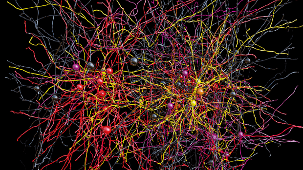
A new browsable 3D image has been created after putting together 225 million images that take up to 1.4 petabytes of data and show one millionth of the cerebral cortex. While it sounds like just one part of the brain is mapped, it still is a huge reveal given how complex our brain actually is.
The human brain is no less than a nexus, and mapping its functions isn’t an easy feat; however, the combined efforts of Harvard and Google pictures the connecting nerves behind the brain’s functions for the first time ever.
Our brain consists of approximately 86 billion neurons connecting through 100 trillion synapses. It is a Godly thing to understand each part’s functioning and how do they alter our thoughts, emotions, behavior, and memory. Scientists are giving it their best shot in building an advanced wiring diagram to understand the functioning and utility of our brain and are calling it a connectome.
Aided by the pandemic, many have realized the importance of understanding the complexities of our brain, a large population have had isolation impacts, and the whole of the thought processes of individuals have been seen changed in some instances, and that might be triggered from extensive isolation periods, and living the life, we were not trained for.
Such advancements in understanding the natural build-up of the human supercomputer “the brain” will aid in finding out the answers and cures for many mental illnesses, allowing to help the affected individuals better.
In 2020, researchers at Google combined with Howard Hughes Medical Institute to encompass half of an insect’s full brain. Followed by Google’s combined efforts with Harvard engineers to map a small section of the human brain, which took putting together a total of 225 million pictures to make up one 3D image. Google is progressing forward in studying the brain and its functions. One of the outcomes of these efforts would be better understanding the brainwaves, which would aid in finding out ways to cure communication between various regions of the brain.
Starting off, scientists behind the project took a 1 mm3 sample from the temporal lobe of the human cerebral cortex. In the first steps, it was stained for visual clarity and then was coated in resin to preserve it. It was then cut into 5,300 slices, each measuring 30 nanometers thick. All the slices were imaged using an electron scanning microscope at a 4nm resolution. Out of this exercise, scientists obtained 225-million two-dimensional images, which were stitched together to form a single 3D volume.
As a result of these efforts, scientists obtained the most comprehensive map of the human brain ever created, including 50,000 cells, 130 million synapses, and even cells such as axons, dendrites, myelin, and cilia. The curious ones can find more about what is explored on the H01 dataset available online.
An idea of how complex and big our brain is could be taken from this single image of a small portion of the brain taking up 1.4 petabytes of data, equaling more than a million gigabytes. The rest is yet to be explored. As per Google, “the sample is just one-millionth of the volume of the brain.”
