
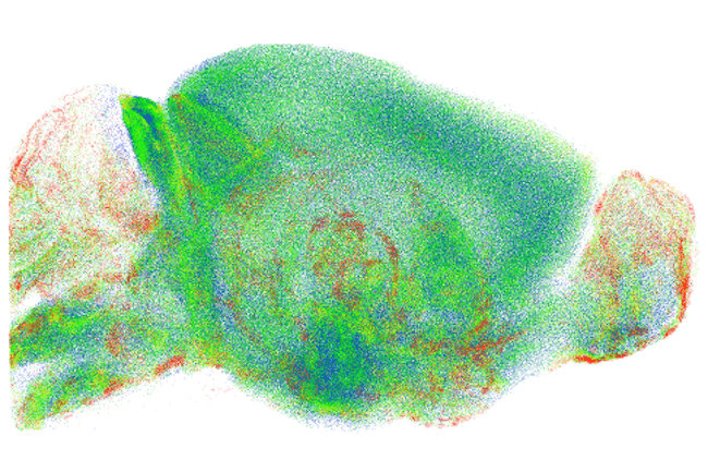
Researchers can now view the mouse brain through the skull, courtesy of a new holographic microscope.
Led by Associate Director Choi Wonshik of the Center for Molecular Spectroscopy and Dynamics within the Institute for Basic Science, Professor Kim Moonseok of The Catholic University of Korea and Professor CHOI Myunghwan of Seoul National University developed a new type of holographic microscope.
The results were published in Science Advances on July 27.
According to the results, it is possible to “see through” the intact skull with this unique microscope. It can provide high-resolution 3D imaging of the neural network within a living mouse brain without removing the skull.
It is important to precisely assess the signal reflected from the target tissue and apply enough light energy to the sample to explore the interior properties of living organisms using light.
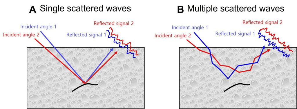
Light experiences repeated scattering in intricate structures like living tissue, which causes photons to erratically shift their direction several times as they pass through the tissue. A large portion of the image data carried by the light is damaged in this process.
By correcting the wavefront distortion of the light reflected from the object to be examined, it is feasible to see the relatively deep characteristics inside the tissues, even if there is only a minimal amount of reflected light. It is vital to reduce the ratio of the multiple-scattered waves and raise it to acquire a high-resolution deep-tissue image.
Specifically, the researchers devised a method to preferentially select single-scattered waves by taking advantage of the fact that they have similar reflection waveforms even when light is input from various angles.
“When we first observed the optical resonance of complex media, our work received great attention from academia,” said Professor KIM Moonseok and Dr. JO Yonghyeon, who have developed the foundation of the holographic microscope.
“From basic principles to practical application of observing the neural network beneath the mouse skull, we have opened a new way for brain neuroimaging convergent technology by combining the efforts of talented people in physics, life, and brain science.”
