
A new revolutionary imaging technique that uses X-rays obtained by a new particle accelerator to produce 3D images of entire organs in high resolution is now available in detail.
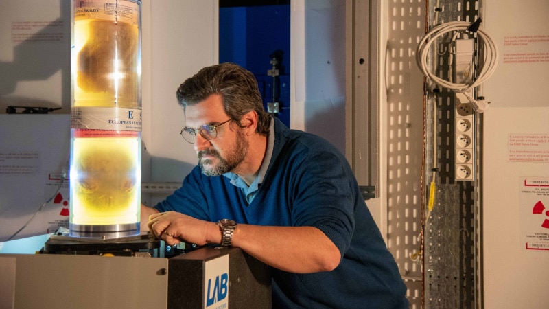
Scientists from the University College London in the United Kingdom and the European Synchrotron Research Facility in France used the world’s brightest X-ray machine, the Hierarchical Phase-Contrast Tomography (HiP-CT), to scan donated human organs, including lungs from a deceased COVID-19 patient.
HiP-CT enables researchers to use 3D mapping of the scanned object at various scales by imaging the entire organ and then zooming down to a cellular level, according to a release.
The technique uses X-rays from the European Synchrotron, which now has the world’s brightest source of X-rays, 100 billion times brighter than a hospital X-ray, kudos to its recent Extremely Brilliant Source upgrade (ESRF-EBS). The brightness enables researchers to see blood vessels five microns in diameter in an intact human lung – about a tenth of the diameter of a strand of hair.
“The idea to develop this new HiP-CT technique came after the beginning of the global pandemic, by combining several techniques that were used at the ESRF to image large fossils, and using the increased sensitivity of the new Extremely Brilliant Source at the ESRF, ESRF-EBS,” explains lead scientist at ESRF Paul Tafforeau. “This allows us to see in 3D the incredibly small vessels within a complete human organ, enabling us to distinguish in 3D a blood vessel from the surrounding tissue, and even to observe some specific cells.”
The HiP-CT is now being used by researchers at University College London to create a “Human Organ Atlas,” released on Thursday. It will show the brain, lung, heart, two kidneys, a spleen, and the lung of a COVID-19 patient, as well as a lung biopsy and a COVID-19 lung biopsy. According to Peter Lee, the project’s leader, the Human Organ Atlas aims to fill a significant gap in our understanding of human anatomy.
The Atlas will be available online to surgeons, clinicians, and the general public.
“Clinical CT and MRI scans can resolve down to just below a millimeter, whilst histology (studying cells/biopsy slices under a microscope), electron microscopy (which uses an electron beam to generate images) and other similar techniques resolve structures with sub-micron accuracy, but only on small biopsies of tissue from an organ,” says Lee. “HiP-CT bridges these scales in 3D, imaging whole organs to provide new insights into our biological makeup.”
Since a steep decline in blood oxygenation levels is a primary sign of COVID-19 worsening. With the help of HiP-CT, researchers found out how severe COVID-19 infection “shunts” blood between two separate systems: the capillaries that oxygenate the blood and those that feed the lung tissue itself. According to the study, the cross-linking prevents the patient’s blood from being properly oxygenated, which was previously hypothesized but not proven.
“By combining our molecular methods with the HiP-CT multiscale imaging in lungs affected by COVID-19 pneumonia, we gained a new understanding [of] how shunting between blood vessels in a lung’s two vascular systems occurs in COVID-19 injured lungs and the impact it has on oxygen levels in our circulatory system,” says Danny Jonigk, a researcher at Hannover Medical School.
According to the researchers, the HiP-CT is intended to provide insights into diseases such as cancer and Alzheimer’s. This previously unseen structural data shows how diseases can influence anatomical structures at resolutions as low as one micron.
“The ability to see organs across scales like this will really be revolutionary for medical imaging,” says Walsh. “As we start to link our HiP-CT images to clinical images through AI techniques, we will – for the first time – be able to highly accurately validate ambiguous findings in clinical images.”
The study was published in the journal Nature Methods.
Source: University College London
