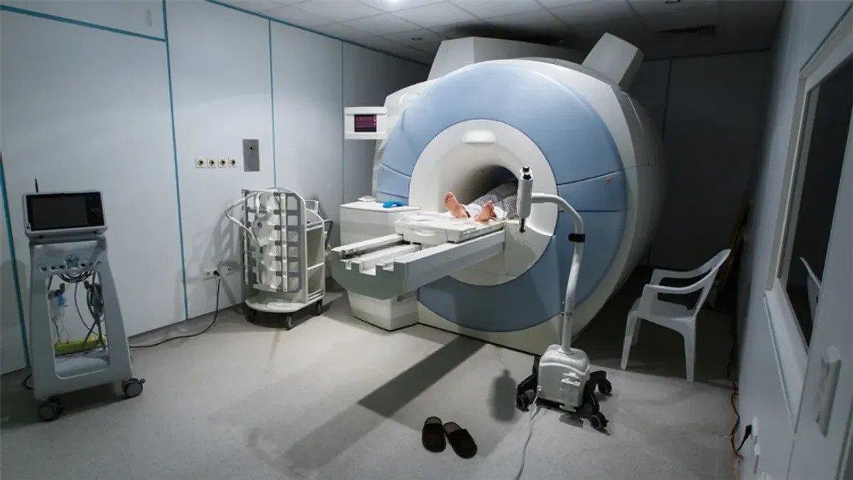
Magnetic Resonance Imaging (MRI) scans, integral to medical diagnostics, enforce a stringent “no metal” policy to ensure patient safety due to the powerful magnetic fields employed in the process. A recent incident, detailed in a US Food and Drug Administration (FDA) report, serves as a stark reminder of the risks associated with disregarding these safety measures.
The report recounts the case of a woman who entered an MRI room with a concealed iron handgun, which, upon exposure to the magnetic field, discharged a single shot into her right buttock.
“The patient was examined by a physician at the site who described the entry and exit holes as very small and superficial, only penetrating subcutaneous tissue,” the report reads, adding that prior to the exam she had been through a standard screening process for magnetic objects “which includes weapons specifically, and answered no to all screening questions”.
Despite undergoing a standard screening process, which includes specific checks for magnetic objects, the patient had falsely denied possession of any. Fortunately, the wounds resulting from the incident were characterized as small and superficial, only penetrating subcutaneous tissue. This event underscores the potentially grave consequences of bringing metal objects, particularly weapons, into proximity with MRI machines.
MRI technology relies on generating powerful magnetic fields and radio waves that target hydrogen nuclei in the body’s water content. The alignment of protons in the magnetic field creates a magnetic vector, and when a radio wave is introduced, the vector is deflected. The emitted signal, produced when the radiofrequency source is deactivated, is then used to create detailed images of internal body structures.
“This uniform alignment creates a magnetic vector oriented along the axis of the MRI scanner,” science editor Abi Berger explains in the BMJ.
“When additional energy (in the form of a radio wave) is added to the magnetic field, the magnetic vector is deflected. The radio wave frequency […] that causes the hydrogen nuclei to resonate is dependent on the element sought (hydrogen in this case) and the strength of the magnetic field.”
“When the radiofrequency source is switched off the magnetic vector returns to its resting state, and this causes a signal (also a radio wave) to be emitted. It is this signal which is used to create the MR images.”
While MRI scans are invaluable for medical diagnostics, the incident emphasizes the critical importance of adhering to safety guidelines. Previous accidents involving MRI machines have occurred when patients introduced metal objects, such as firearms, leading to serious injuries or even fatalities.
This underscores the need for comprehensive education regarding the potential dangers and the strict enforcement of safety protocols to prevent accidents during medical procedures involving MRI scans. Ultimately, the incident serves as a cautionary tale about the severe consequences that can arise from negligence in adhering to established safety measures surrounding MRI technology.

