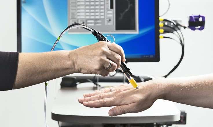
Although Medical Science and Surgery have improved beyond recognition, Cancer surgery still uses the gut of the operating surgeon more than anything else to determine the extent of the cancerous region. They don’t have a way to go molecular at that stage and find out the exact cancerous points on the affected area. Many experienced surgeons can locate tumors due to a plethora of experience under their belt, but they reach that stage after several setbacks on live patients where they cut off something that wasn’t cancerous and have to carry the weight of the guilt with them. This new pen microscope aims to help them locate the exactly affected area using a new microscopy technique called dual-axis confocal microscopy.
Developers from the University of Washington, who were behind the realization of this project, believed that time had come to make an accurate device that can allow doctors to follow the path of cancer. They always have to rely on professional instinct which is not very accurate and leads to disasters now and then. With this pen microscope, they can zoom in and out at the tissue ahead of them and thus find out the exactly affected regions without a need for guessing games.

The problem with microscopes was that there was no handheld device that could take us to this level and help spot the cancer overgrowth. The pen can not only magnify but also help map these areas and even light up tissue parts more than half a millimeter beneath the visual surface. With this multi-faceted observation platform, doctors will have a much better understanding where to cut or not in a Cancer operation. According to the lead researcher Jonathon Liu, working with cancerous tissue is like driving in thick fog. Their experience might be able to get you somewhere, but it is not a guarantee you won’t bump into the car in front of you!


