


Scientists have developed an advanced new brain sensor that will be able to record electrical signals from the brain’s surface in record-breaking resolution, which can help neurosurgeons better differentiate between healthy and diseased tissue and deepen our understanding of how the human brain functions.
It has been brought by the engineers at the University of California (UC) San Diego and is what is known as an electrocorticography (ECoG) sensor. These sensors are commonly placed on the exposed brain cortex during surgery to record the emanating electrical signals and reveal which regions of the brain tissue are active. This can in turn enable neurosurgeons to safely remove brain tumors or sections of tissue where epileptic seizures are originating while leaving healthy tissue untouched.

This will lead to an improvement in the preservation of healthy, functioning brain tissue, and this is the goal being pursued by the UC San Diego researchers. The ECoG devices in use today are mostly comprised of somewhere between 16 and 64 sensors, though some research-grade examples can include up to 256. The UC San Diego team was able to produce ECoG grids with either 1,024 or 2,048 sensors, through some innovative engineering breakthroughs.
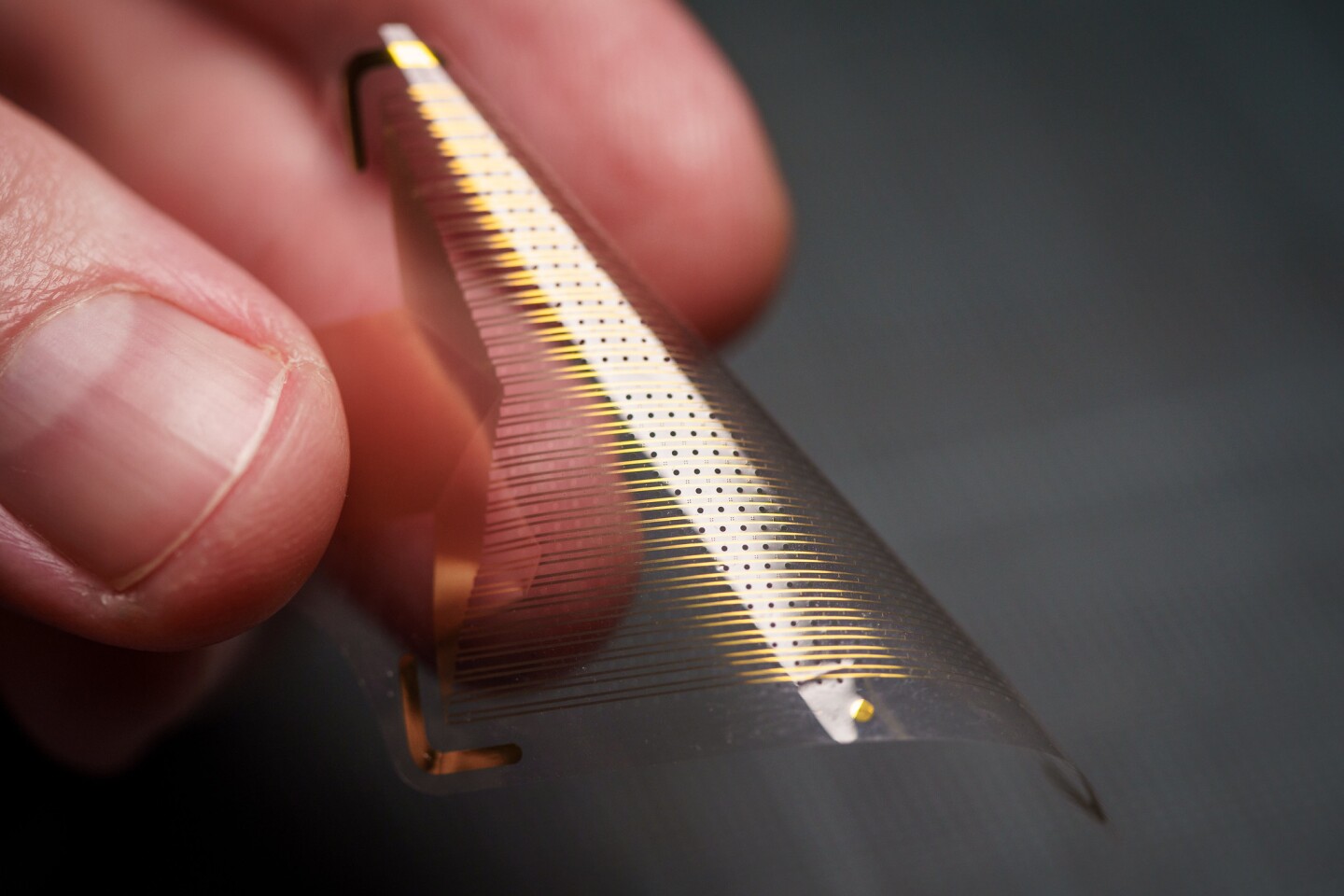
Clinically approved ECoG grids feature sensors are spaced a centimeter (0.4 in) apart, to avoid interference between them. The team used nanoscale platinum rods, which offer a greater sensing surface area than the flat platinum sensors used today and allow for placement of 100 sensors per unit area instead of one, or 100 times the spatial resolution.
These rods are placed one millimeter apart on a flexible, biocompatible material called parylene, with the resulting sensor grids around 100 times thinner than those used today, at around 10 micrometers thick or around one-tenth the size of a human hair.

They experimented by using the 1024-sensor version to directly record signals from the brain tissue of 19 patients undergoing surgery. They also used the sensors to map key regions of the brain in four different subjects during motor tasks and used them to map the cortical column of a rat brain for the first time.
The team is now aiming to acquire the clinical approval of the technology so it can help the sufferers of treatment-resistant epilepsy and brain tumors
The team also explored the areas like mind control of prosthetic limbs, treating memory loss, and interacting with the digital world without the need for smartphones by using the sensors to monitor brain activity associated with finger sensation and hand grasping.
The research was published in Science Translational Medicine.