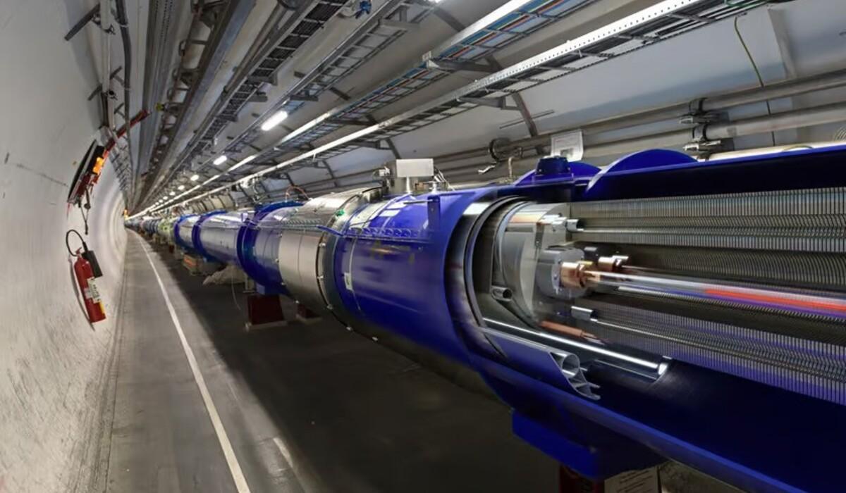
In Germany, physicists are repurposing technology initially developed for particle accelerators at CERN to enhance the precision and safety of brain tumor treatments. Traditional methods for destroying head and neck tumors often pose risks to healthy tissue. Ion beams, accelerated to three-quarters the speed of light, can penetrate tissue deeply but pose a risk of damaging surrounding healthy cells, especially in delicate areas like the brain.
To address this challenge, researchers are utilizing a novel imaging device incorporating the Timepix3 pixel detector, originally developed at CERN. This technology allows for precise tracking of secondary radiation emitted by the ion beam, updating tissue maps in real-time. By comparing the emitted radiation to the initial CT scan, any shifts in the brain’s position within the skull can be accounted for during treatment.
The imaging device, developed by ADVACAM, registers each charged particle emitted from the patient’s body, akin to tracking billiard balls after a shot. If the observed particles align with the CT image, the treatment is confirmed on target. However, discrepancies prompt adjustments to ensure accurate targeting of the tumor. These adjustments aim to minimize unnecessary radiation exposure to healthy tissue while maximizing radiation delivery to the tumor.
Currently, treatment adjustments necessitate interrupting the procedure for replanning. However, future iterations aim to enable real-time corrections to the ion beam’s path during treatment. This advancement holds promise for more efficient and effective tumor targeting while reducing the risk of collateral damage.
The adaptation of CERN-developed technology for medical applications underscores the versatility of scientific innovations. Originally designed for particle physics research, the Timepix3 detector now finds application in healthcare, demonstrating the interdisciplinary nature of scientific progress. Michael Campbell, spokesperson for the Medipix Collaborations, emphasizes the unforeseen yet impactful applications stemming from collaborative research efforts across diverse fields.
“Our cameras can register every charged particle of secondary radiation emitted from the patient’s body,” said Lukáš Marek from ADVACAM. “It’s like watching balls scattered by a billiards shot. If the balls bounce as expected according to the CT image, we can be sure we are targeting correctly. Otherwise, it’s clear that the ‘map’ no longer applies. Then it is necessary to replan the treatment.”
The idea is that these updates will better target the tumor while reducing the amount of unwanted radiation the patient is exposed to while hitting the tumor with higher levels of radiation.
At the moment, the detector requires interrupting the treatment to allow for replanning. However, later phases of the program will include the ability to correct the beam path in real time.
“When we started developing pixel detectors for the LHC we had one target in mind – to detect and image each particle interaction and thereby help physicists to unravel the secrets of Nature at high energies,” says Michael Campbell, Spokesperson of the Medipix Collaborations. “The Timepix detectors were developed by the multidisciplinary Medipix Collaborations whose aims are to take the same technology to new fields. Many of those fields were completely unforeseen at the beginning and this application is a brilliant example of that.”

