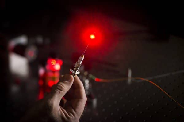
Researchers from University College London (UCL) and Queen Mary University of London (QMUL) have collaborated on a project to develop a new optical ultrasound needle. This surgical needle allows heart tissue to be imaged in real-time during keyhole procedures.
The surgical needle has been successfully tested on pigs and gives the doctors a high-resolution image of soft heart tissue up to 2.5 cm in front of the needle. The needle has the potential to make surgery not only faster but more accurate as well. Doctors have to rely on preoperative imaging scans and external ultrasound probes to help them visualize the soft tissue that was being operated upon during a keyhole surgery.
The hole is too small to add imaging devices as well, and this breakthrough technology is a major game changer. Dr. Malcolm Finlay, study co-lead and consultant cardiologist at QMUL and Barts Heart Centre describes the new hardware, “The optical ultrasound needle is perfect for procedures where there is a small tissue target that is hard to see during keyhole surgery using current methods and missing it could have disastrous consequences. We now have real-time imaging that allows us to differentiate between tissues at a remarkable depth, helping to guide the highest risk moments of these procedures. This will reduce the chances of complications occurring during routine but skilled procedures such as ablation procedures in the heart. The technology has been designed to be completely compatible with MRI and other current methods, so it could also be used during brain or fetal surgery, or with guiding epidural needles.”
The tool works by putting a miniature optical fiber inside a specially designed surgical needle. The needle can deliver pulses of light that generate ultrasonic pulses. These pulses are reflected from the soft tissue and are detected by a second optical fiber equipped with a sensor that provides real-time ultrasound imaging.
The study’s co-author, Dr. Richard Colchester (UCL Medical Physics & Biomedical Engineering) describes the new tool’s application: “The whole process happens extremely quickly, giving an unprecedented real-time view of soft tissue. It provides doctors with a live image with a resolution of 64 microns, which is the equivalent of only nine red blood cells, and its fantastic sensitivity allows us to readily differentiate soft tissues.”
The technology has only been made possible by the development of a black material that contains a mesh of carbon nanotubes and the creation of optical fibers based on polymer optical microresonators for detecting ultrasound waves. The team is still working together and might use this technology to develop other keyhole surgery tools.

