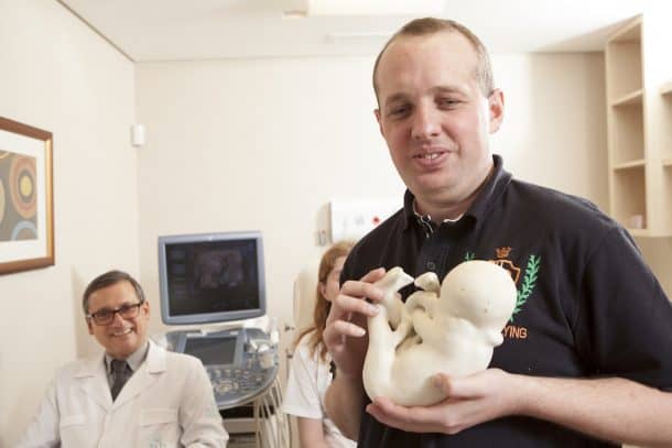
3D printing can help us in a lot of ways, and that is not limited to manufacturing or health tech. For a parent, seeing the first ultrasound of the baby is one of the most beautiful and exciting moments, when a parent gets to bond with their unborn child seeing the sonogram and hear its little heart beat. A visually impaired person, however, is deprived of that wonderful feeling. Here is the point where 3D printed ultrasound comes into play.
In 2014, a legally blind Brazilian couple, Ana Paula Silveira and her husband Alvaro Zermiani went in to get their first ultrasound to check on the health of their developing fetus, knowing they could not get to experience the joy of actually looking at the baby breathe. The couple was overjoyed when they got to hold a 3D printed replica of their unborn child. Instead of relying on others for a description of the fetus, they could feel it, its face, and features.
GE Healthcare brought in the 3D printed ultrasound technology from Voluson E10. The source from where the idea sprang is the most expected of all. Fossils, which are the exact opposites of babies, brought the idea as Dr. Heron Werner, a Brazilian obstetrician, and gynecologist saw a 3D printing project at the National Museum of Brazil that digitized ancient exhibits. The technology inspired Dr. Werner to bring the same technology to fetuses, and he began to give visually impaired parents, 3D printed ultrasounds free of charge.
Ana Paula had seen a TV interview with Dr. Werner and later decided to approach him for help with a 3D sonogram when she became pregnant in 2013. The child is now a healthy three-year-old named Davi Lucas Zermiani who now gets to play with the 3D model of his fetus self.

Voluson E10 is the first machine in the field of obstetrics/gynecology that also allows direct 3D printing from the computer. Even though the idea was to enable visually impaired people to feel it but all parents could keep it as a sentimental memory of their experience. The technology even allows a better detection of health problems in the fetus than a traditional sonogram, finding out the tiniest of details like cleft lips, abdominal wall defects, or any other physical abnormalities. They can even serve as educational tools for better surgical planning.


