
Wellcome Image Awards 2017 is here, and like every year, it is sure to blow your minds! The finalists of this year have been announced, along with the display of some breathtaking scientific imagery that is nothing short of art!
Here are our favorites:
Vessels in a Pig Eye
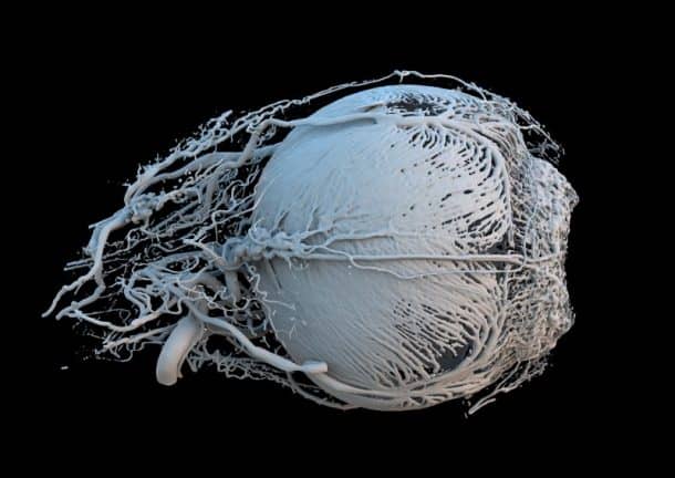
The researchers from Switzerland used incredibly accurate computed tomography (CT) and 3D printing to create this image which depicts the blood vessels within a pig’s eye. The vessels bring energy and food to the muscles surrounding the iris.
Surface of a Mouse Retina
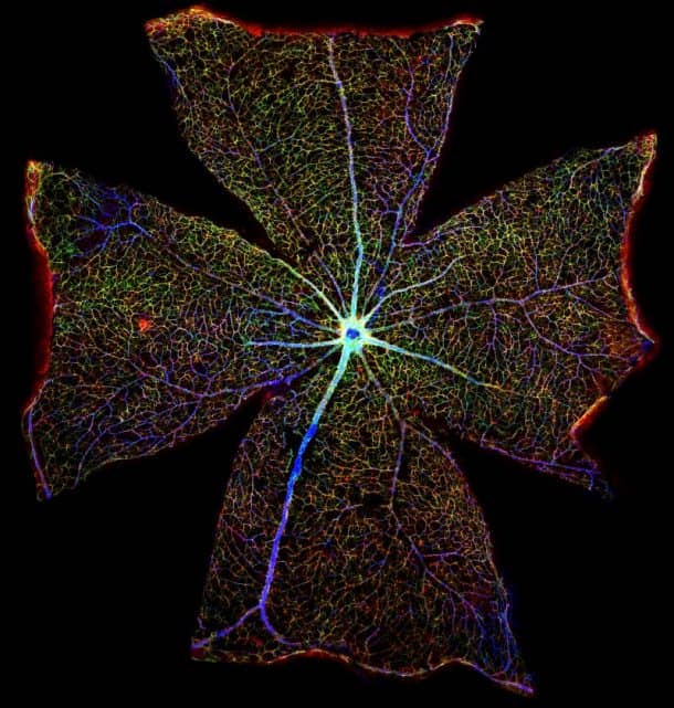
Almost 400 microscopic images were stitched together to make the magnificent picture of a mouse’s retina. The blue lines are blood vessels, and astrocytes are visible in red and green.
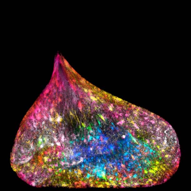
The Placenta Rainbow

These pictures of placentas are taken from genetically modified mice, and each has a distinct immune system. The “Placenta Rainbow” is the result of the differences in placental development, rising from the manipulation of the female mice which was done in a bid to understand and eventually, treat problems that arise during human pregnancies.
Unraveled DNA in a Human Lung Cell
Believe it or not; this is the nucleus of a human lung cell packed with DNA. The deformed shape of the cell results from tension caused by rope-like strands of DNA pulling between the two cells.
Zebrafish Eye
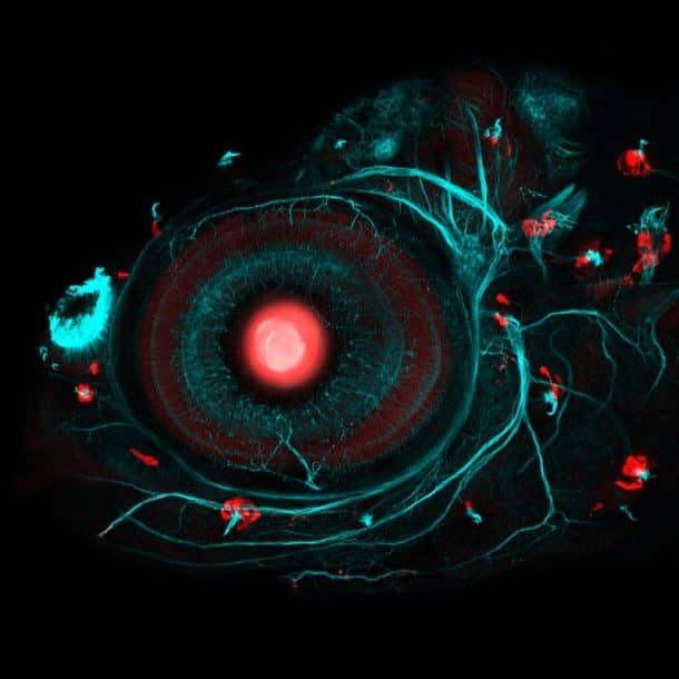
The picture above shows the eye of a four-day-old embryo zebrafish. The researchers from the University of College London used CRISPR-Cas9 gene-editing and strategic breeding to create a zebrafish that had its body parts glow in fluorescent red.
Cat Skin
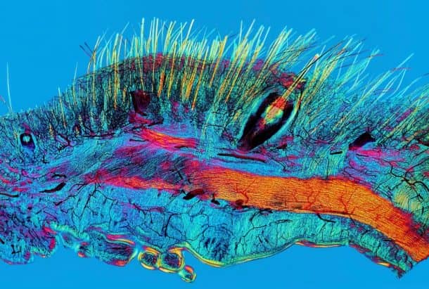
This picture of a cat’s skin was taken via polarized light micrograph, which only allows light traveling in a particular orientation to pass. Yellow strands are hairs and whiskers while the blood supply can be seen in black.
Iris Clip
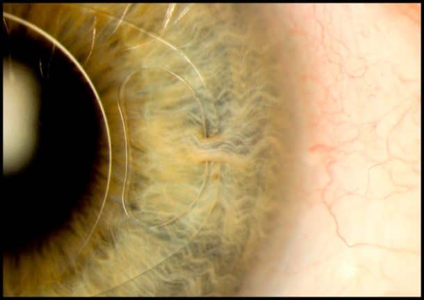
The “iris clip” is used to treat nearsightedness and cataracts, and the high-definition shot above shows how the artificial intraocular lens (IOL) is fitted onto the eye via the small surgical incision.
Blood Vessels of an African Gray Parrot
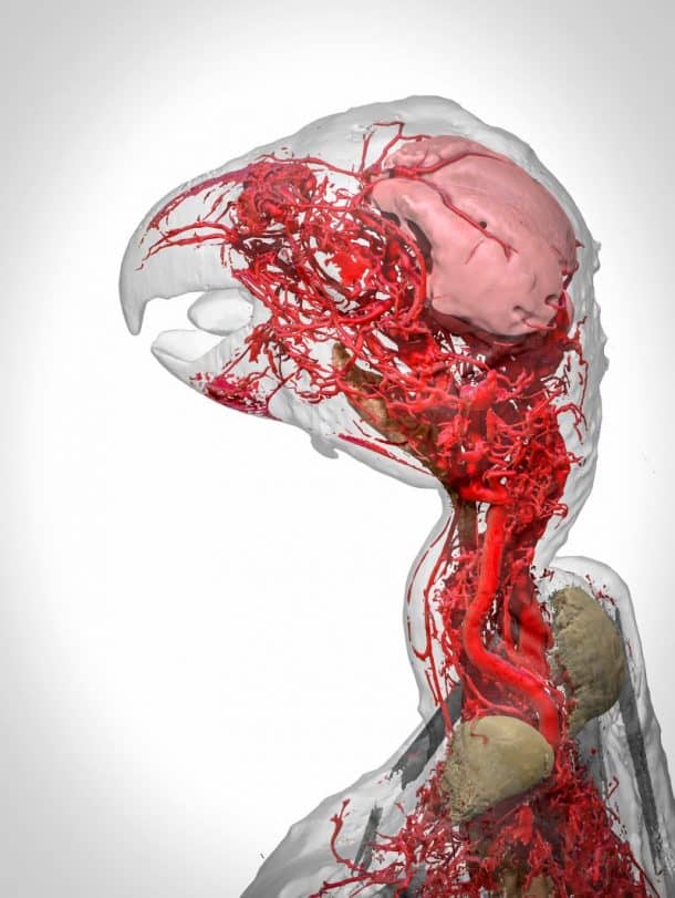
The image above is a 3D reconstruction of an African gray parrot created using a new contrast agent called BriteVul. The model highlights the intricate system of blood vessels in the head and the neck of the bird.
The winners of the contest will be announced on March 15 at the Wellcome Trust in London, and we already feel pity for the poor judges who will have to choose among these masterpieces.
You can view more pictures here.

Soler