

The medical science has improved quite a lot over the last few decades and now we undergo a myriad of operations without a second thought. However, if you were alive before 1846, you would have known the nightmare it was to undergo an operation, without anesthesia! The first pain-numbing drug was employed in 1846 but before that, the patients were operated upon while they were conscious.
A new book that contains images, detailed, of surgical textbooks hailing from the 17th, 18th and 19th centuries has been published. The book is not for the faint of heart since it features brains being slices and eyeballs being pierced. The book has been named as Crucial Inventions and has been drawn from The Wellcome Collection’s library – narrated by medical historian Richard Barnett.
Many patients used to die from the post-operative shock, loss of blood or infection. The death rate after operation was about 80% in some of the hospitals in London. You can see some of the images below from the book. (Warning : NOT for the faint of heart).
Eye surgery to correct ‘strabismus’, a misalignment of the eyes known as a squint.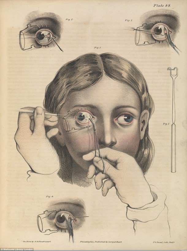

Surgery to remove cancer of the tongue – Tongue was sliced in two (Fig. 1), the tumor was cut out (Fig. 3) and tongue was stitched back together (Fig. 3).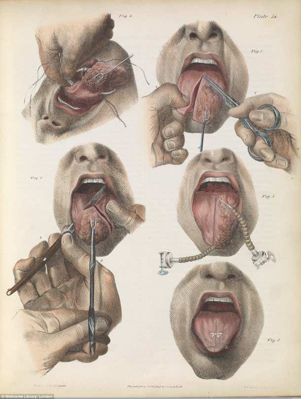

Amputation of toes and foot meant simply cutting them off with a knife (Fig. 1). 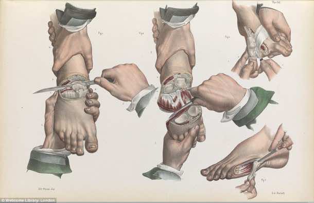

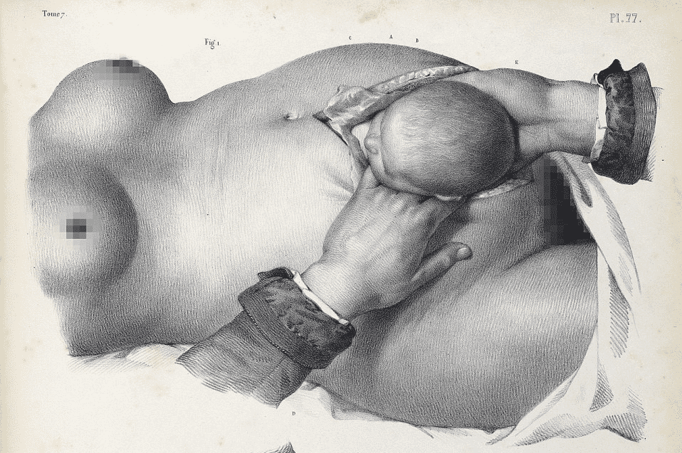
1840 lithograph shows the painful process of having a Caesarean section before anesthetics were used.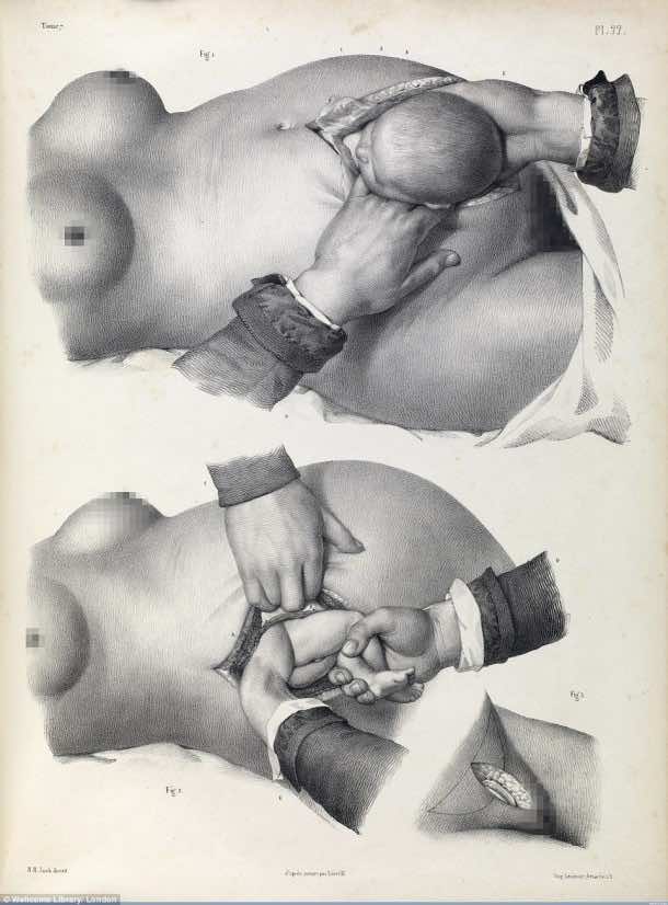

This is from an 1841 textbook and depicts how doctors carried out the removal of breast and dressing of wound afterward. 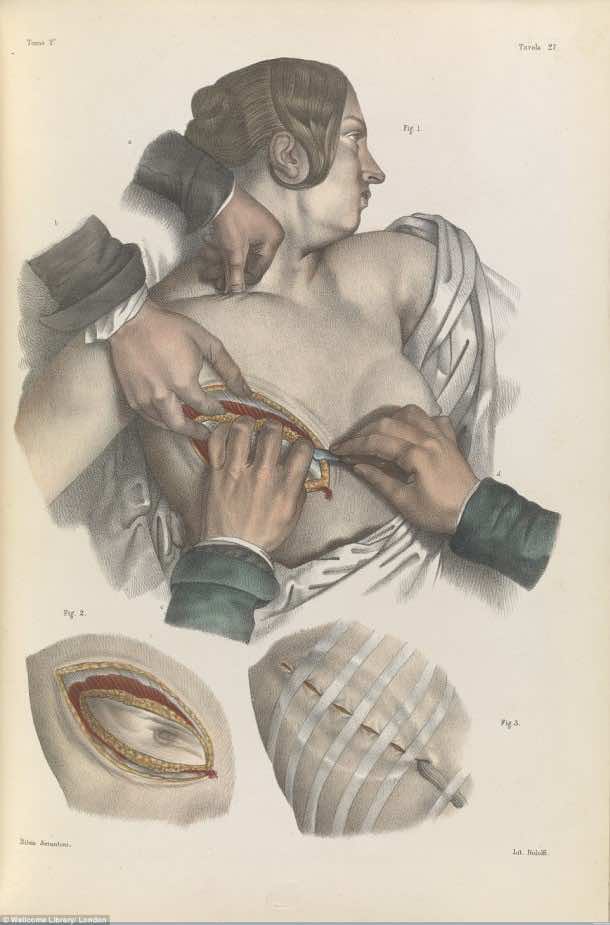

An 1841 surgical book shows how doctors would reconstruct the lower jaw to prevent diseases of the mouth. 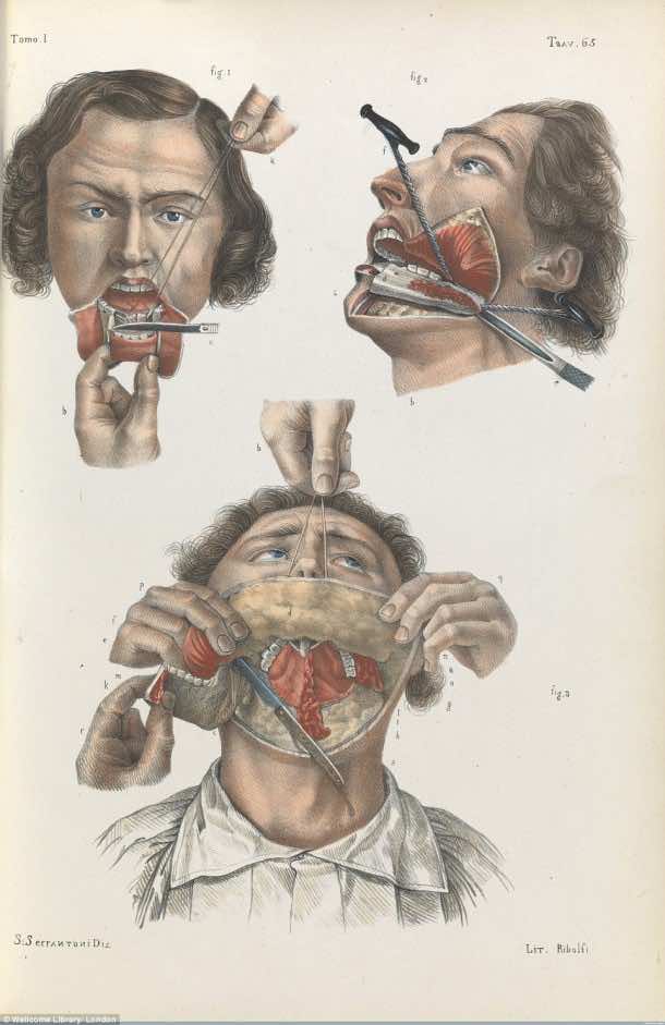

1841 text book shows an artery in the groin being sewn by doctors using sutures and a suture hook, while compressing the abdomen to reduce blood flow.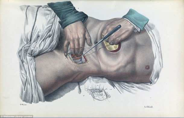

An 1844 image shows a vertical cross-section of the human brain.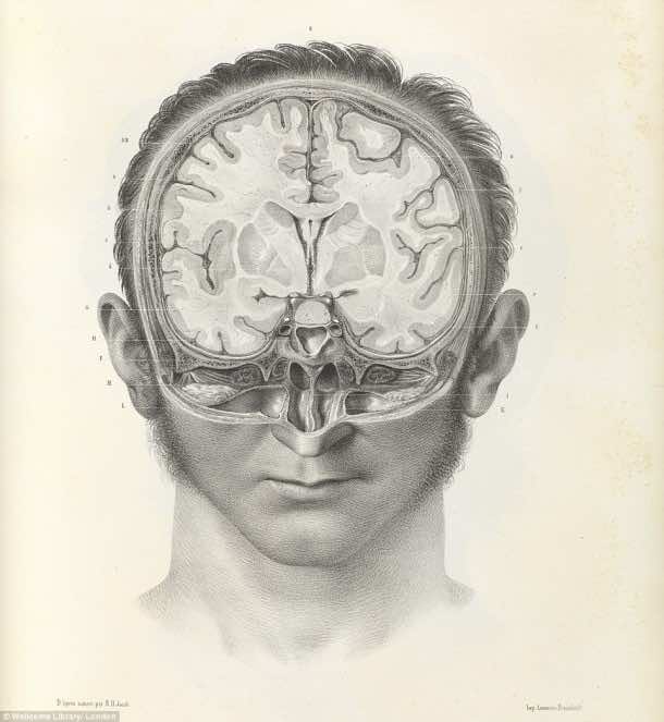

Image from an 1841 textbook shows the barbaric-looking surgical instruments used for amputation, bone and organ operations.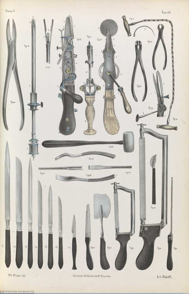

Image from a book, circa 1675, shows treatment for lacrimal fistula (a small lesion near the eye).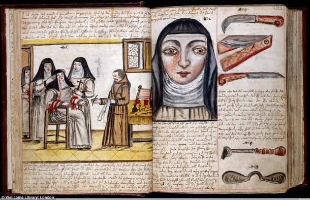

Illustration from an 1846 textbook shows various surgical procedures.

Illustration from an 1866 book shows arteries in the lower arm and elbow being tied up by surgeons to cease blood flow.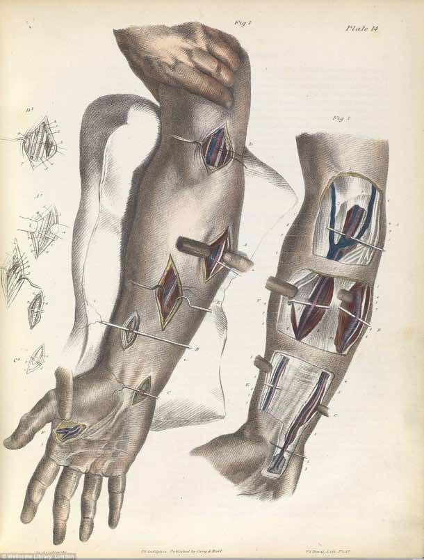

A diagram from an 1848 book shows the anatomy of the armpit, with the axillary artery, the vessel that takes blood to the neck and abdomen, tied off to stop blood flow.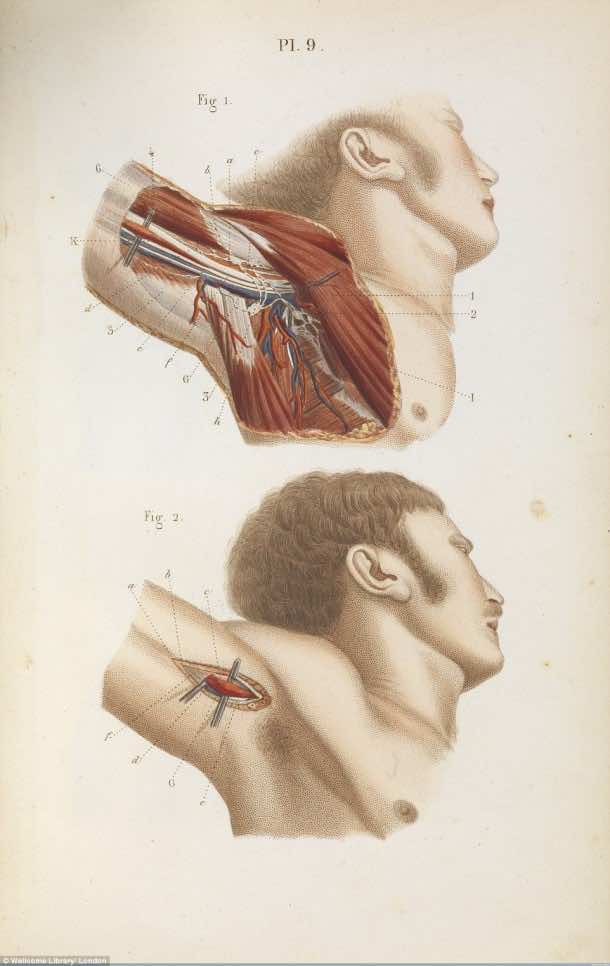

An image from circa 1675 shows a woman’s breast operation – slicing off the organ actually.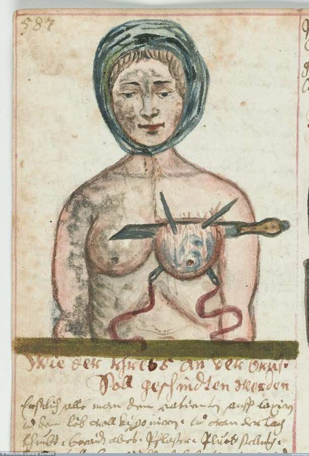

Circa 1675, shows the practice of bloodletting, or withdrawing blood to prevent illness or disease.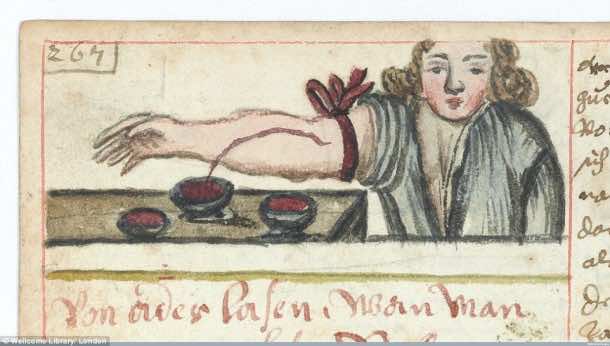

Diagram from a 1856 book shows how doctors would dissect the chest to reveal the lungs, heart and main blood vessels.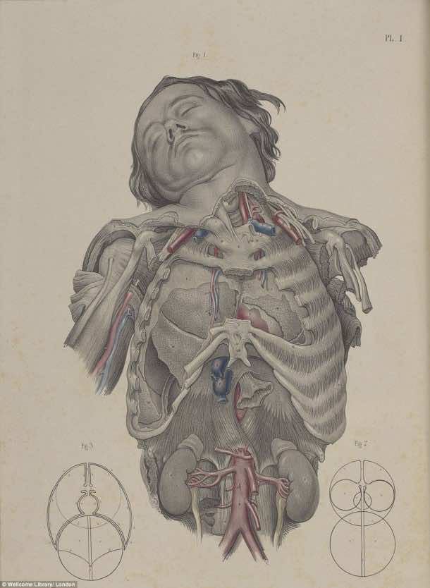

Crucial Interventions or, An Illustrated Treatise on the Principle and Practice of Nineteenth-Century Surgery by Richard Barnett is published by Thames & Hudson, in association with the Wellcome Collection.