Many of us who have taken Biology in high school have experienced using a microscope and it all appears to be a set of obscure minute objects that don’t make sense unlike the bright colored diagrams from out textbooks. But, what we don’t know is that to see anything in a beautiful way, one needs to know how to set the lens and lighting in the right manner. I am not talking about focusing them, as any person can do that. What I mean is that to observe the beauty of something, one must know the lighting, angles, colors and definition required to capture them. It sounds like a job for a photographer and that too who knows about microscopy.
Apparently, microscopic photography isn’t an obscure field as there are many well-trained professionals around the world who do it for a living. An annual event named Nikon Small World Competition brings together some of the finest work from this small world and displays it on their website. It is still going on and winners will be announced by 14th of this month. Seeing the pictures below, the judges will have a tough choice to announce the best photographer at this event of covering toadstools, bees and other creepy crawlers.
Here are some amazing pictures from the finalists:
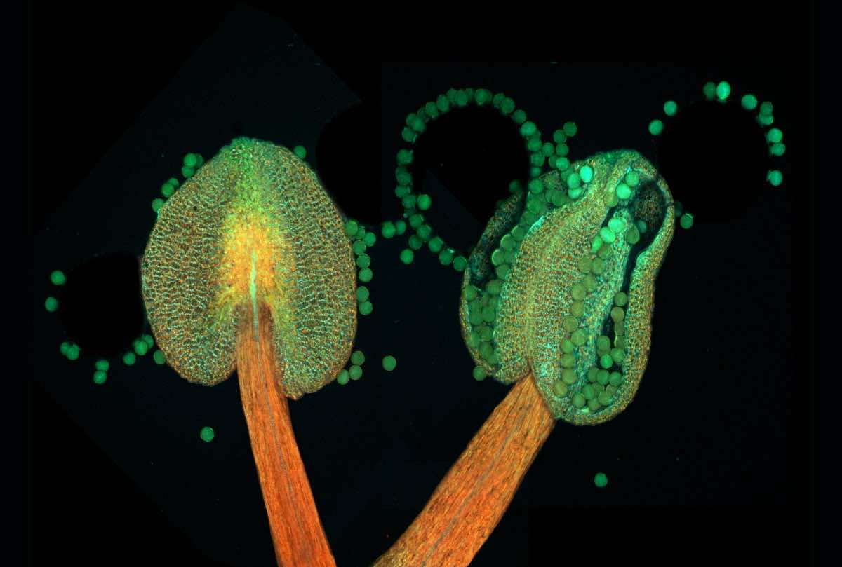
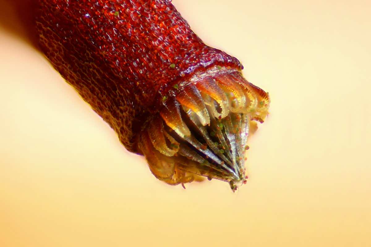
The anther containing pollens
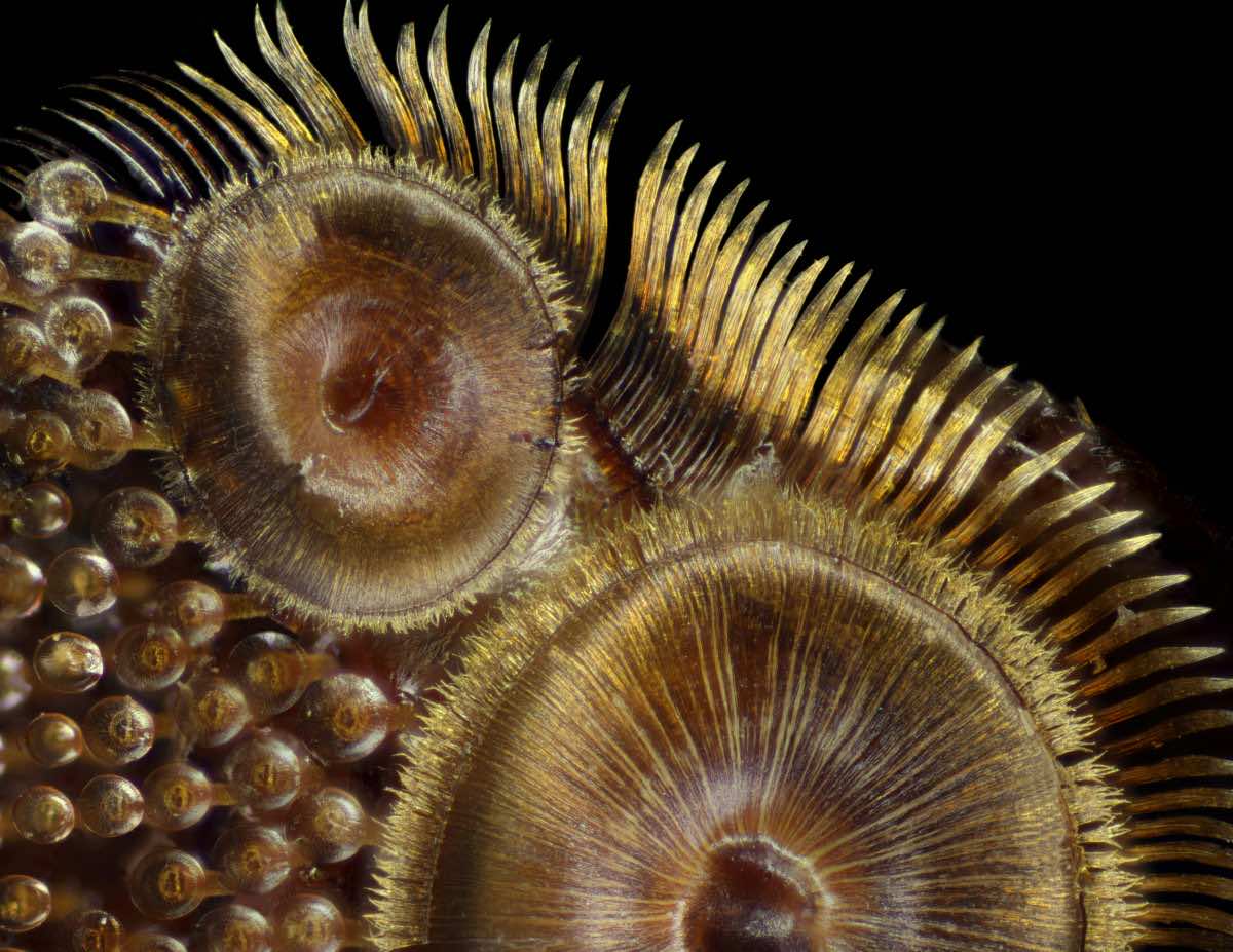
Feeding structure of an aquatic snail
Numerical traces on a Blue-ray disc

Torn photographic film
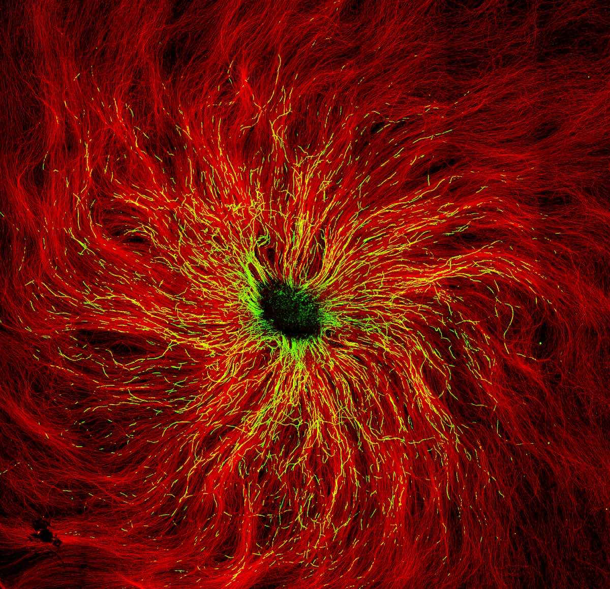
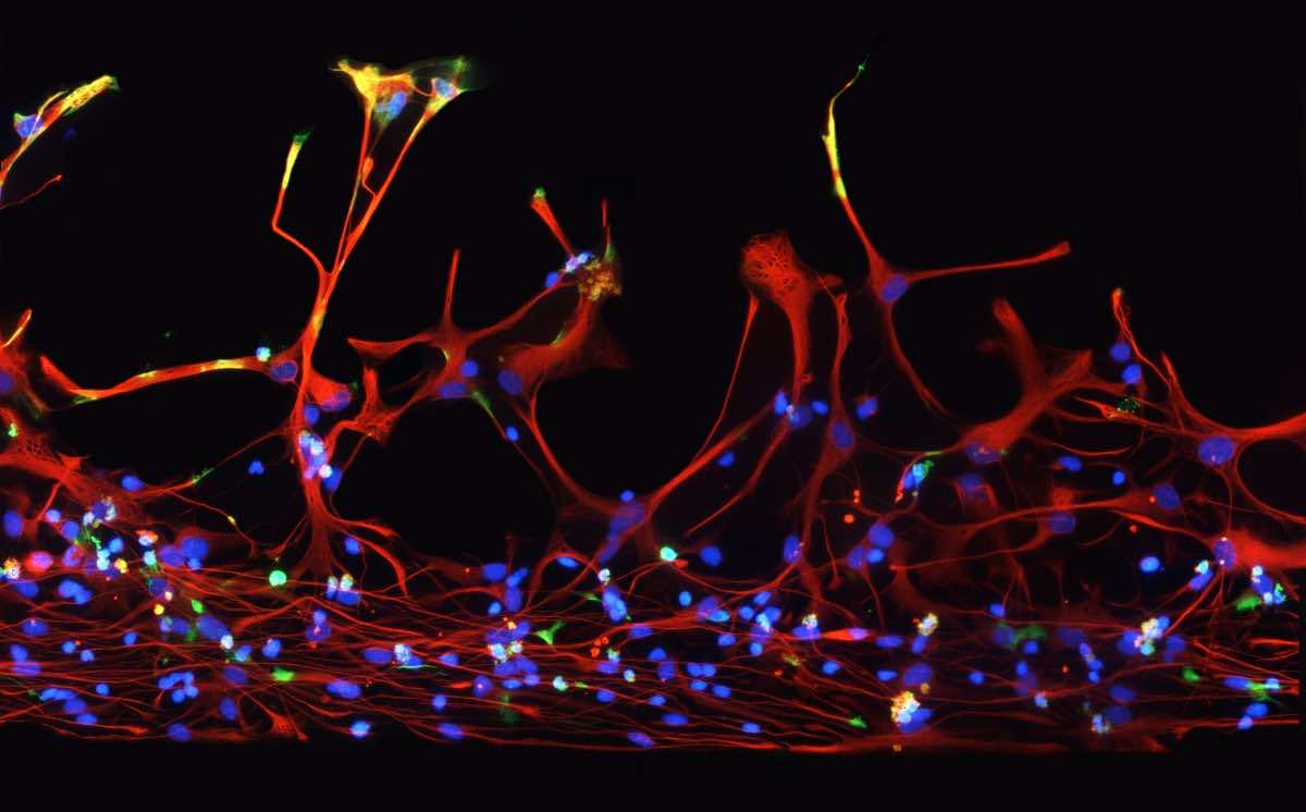
Neurons from mice relaying information
Tentacles of a flesh-eating plant
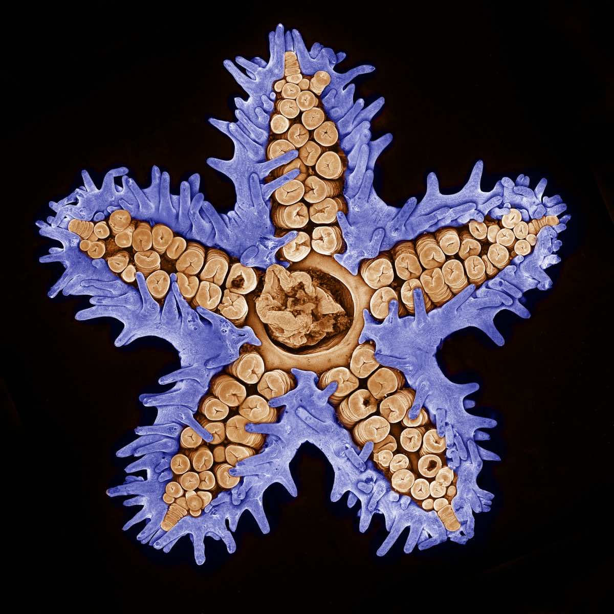
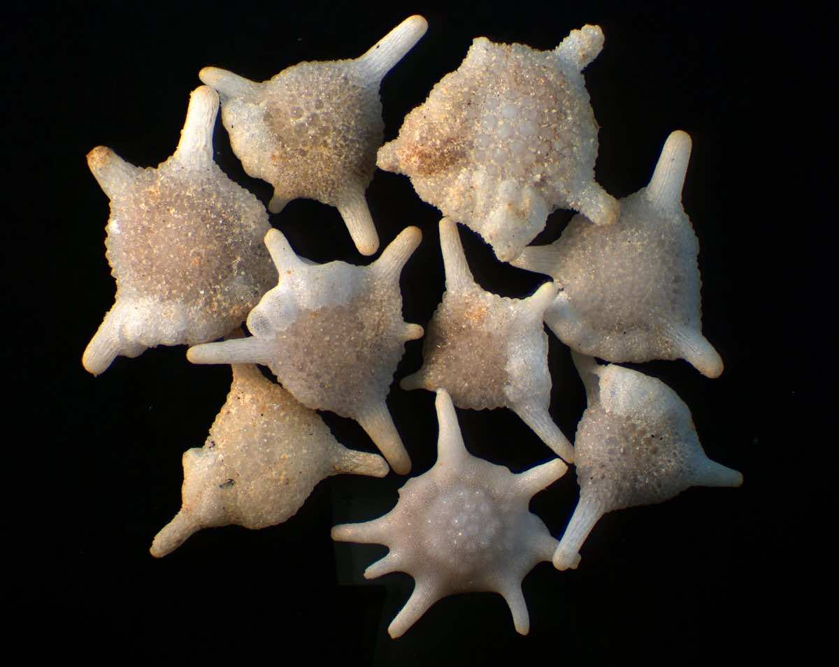
 A young starfish
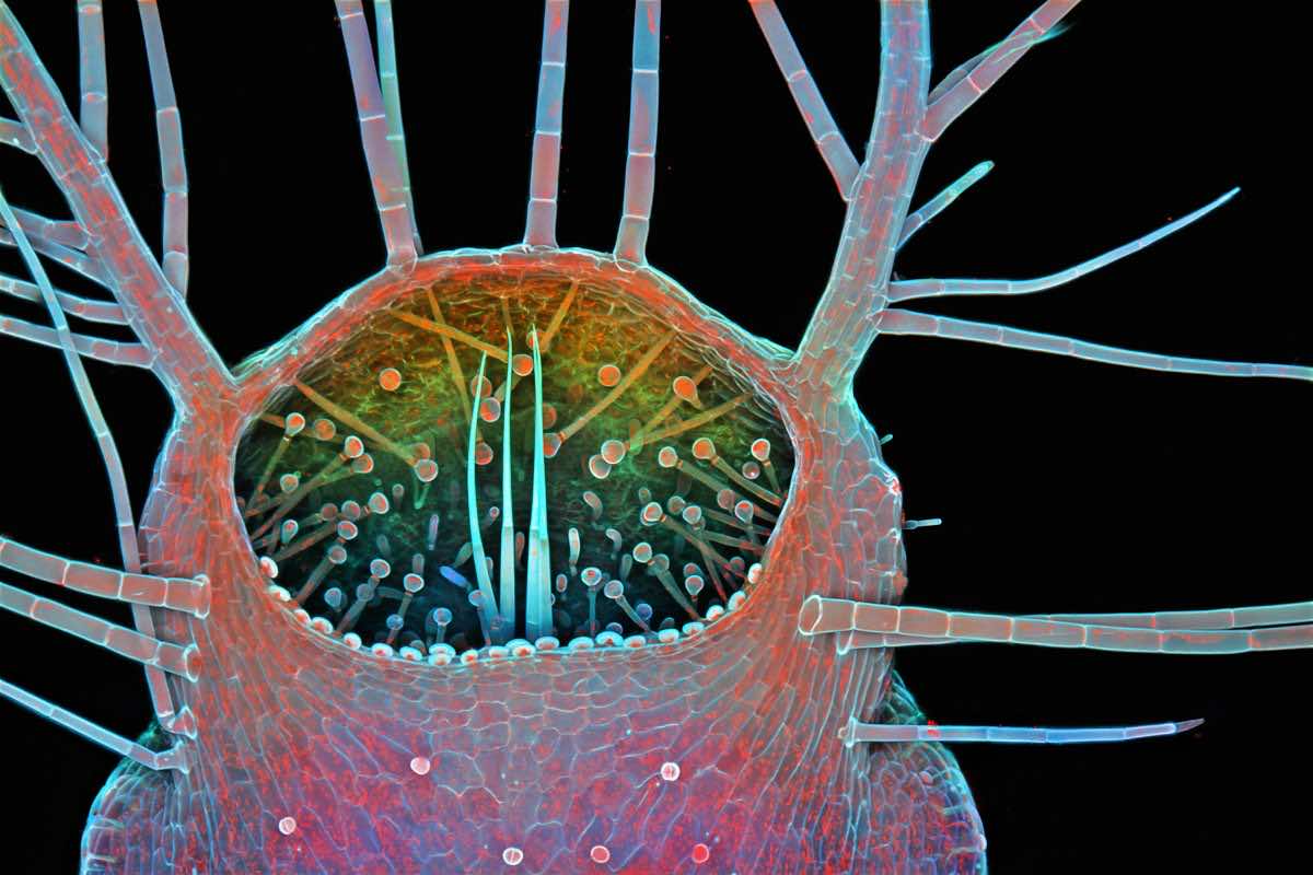
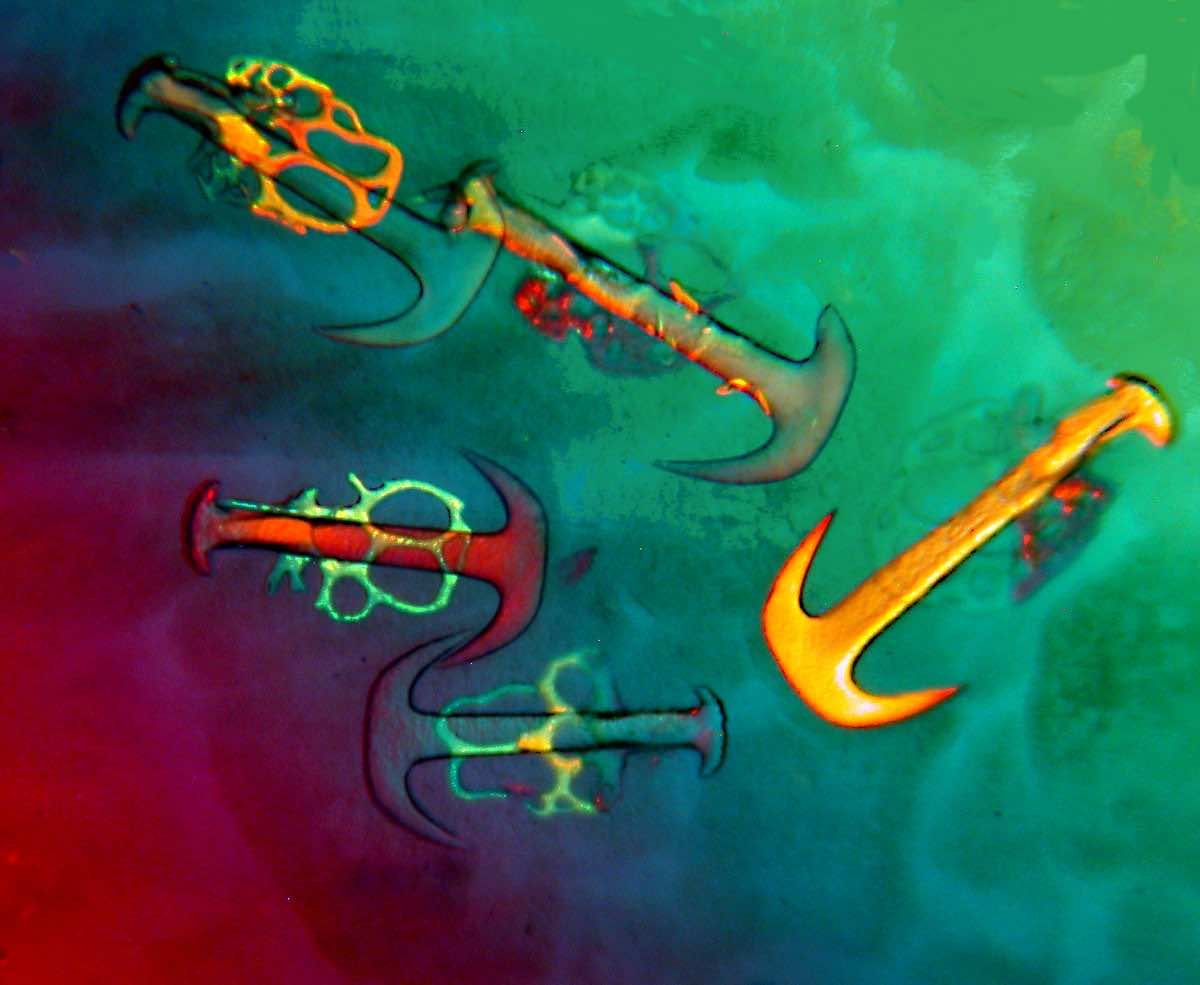
Intake of a young bladderwort
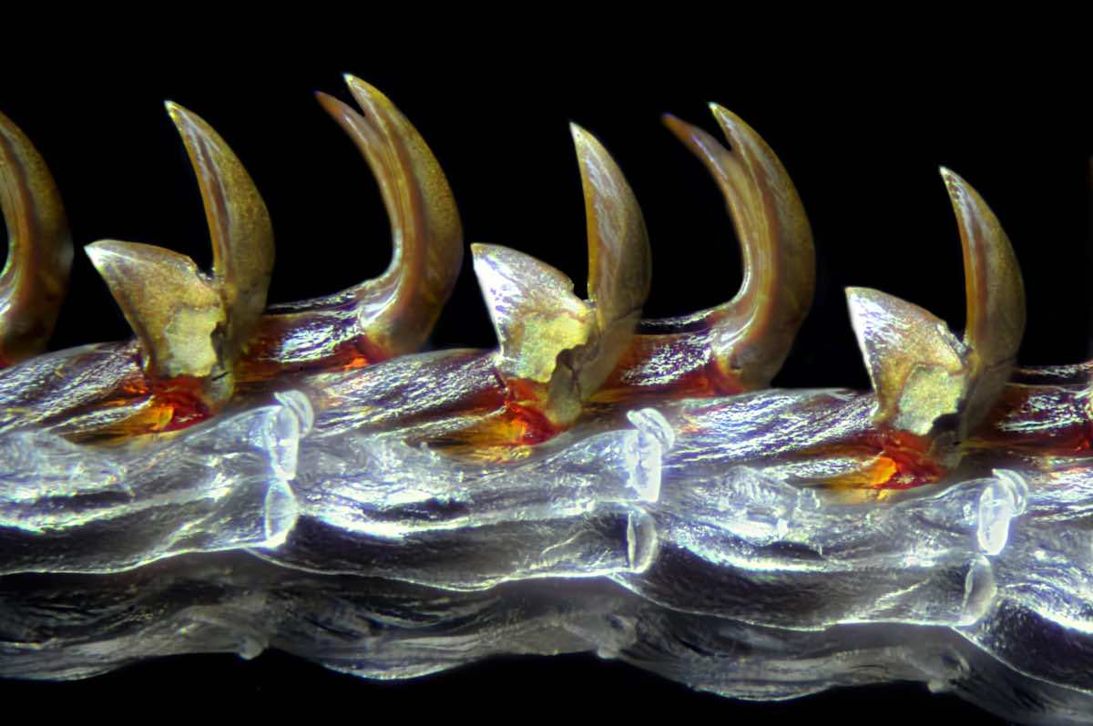
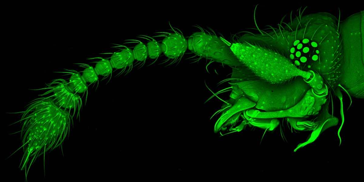
Rove Beetle Head
Spore capsule of a moss
Acne medication in crystallized form
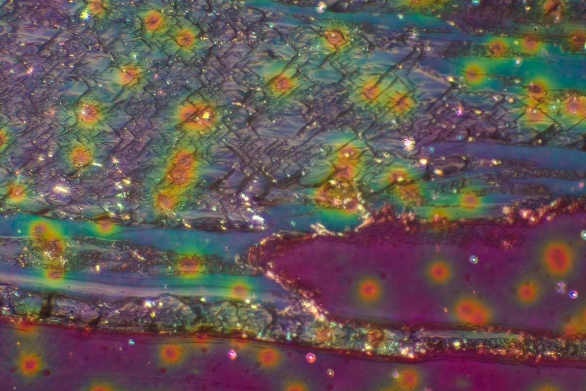
Cross section of a prehistoric horse bone
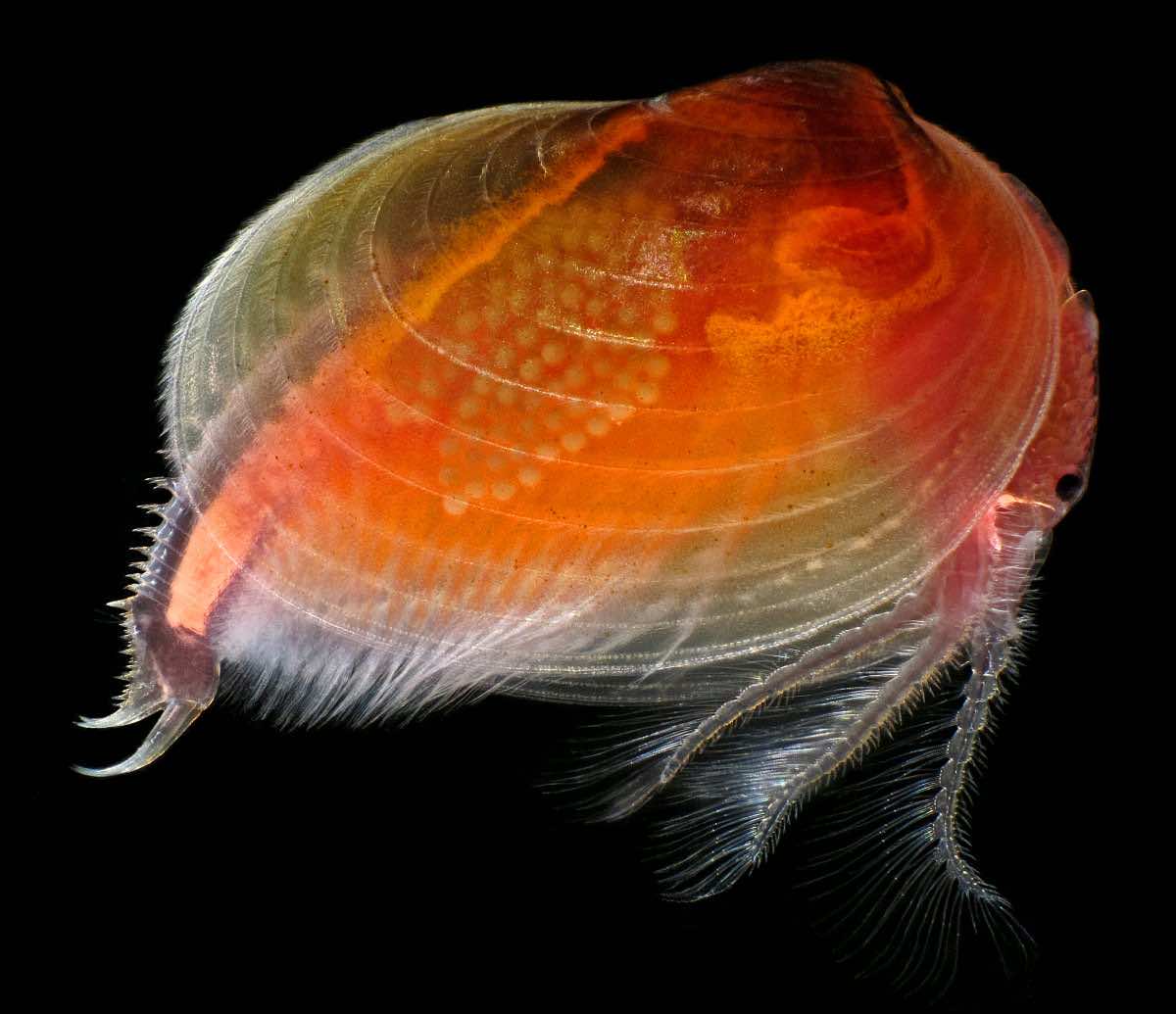
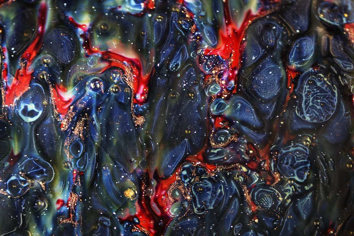
Clam’s different colors
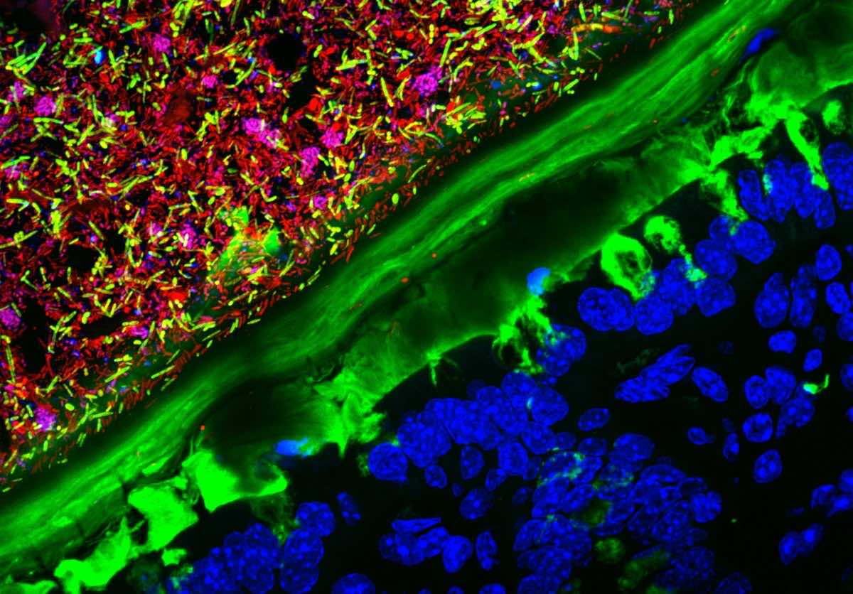
Mouse colon inhabited by human microbes
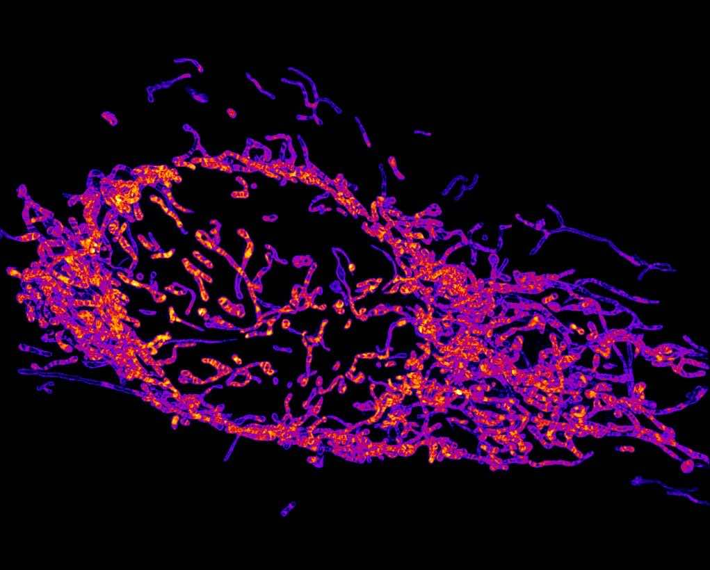
Mitochondrial of a cell
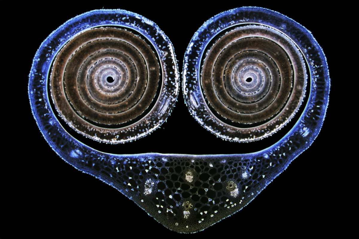
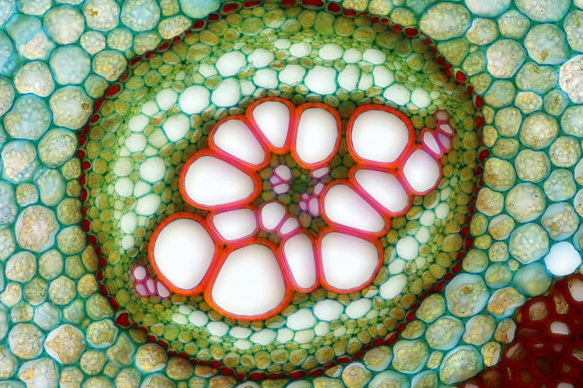
Water Lily bud’s cross section
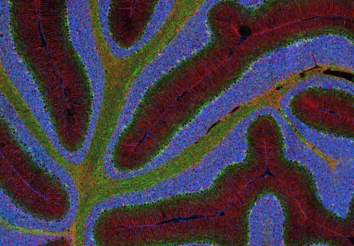
Part of Rat’s brain
Moth’s mouth parts
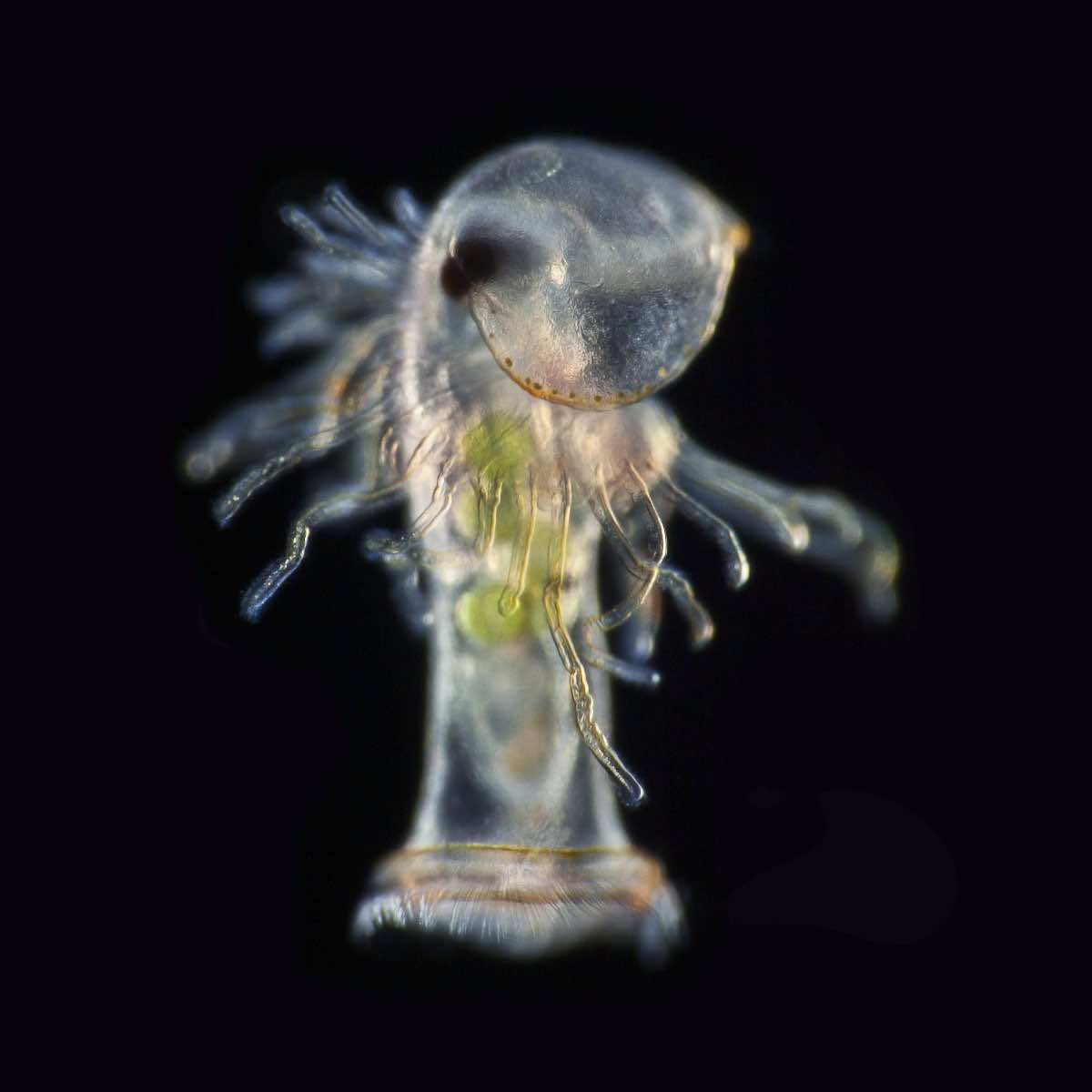
Offspring of horseshoe worm
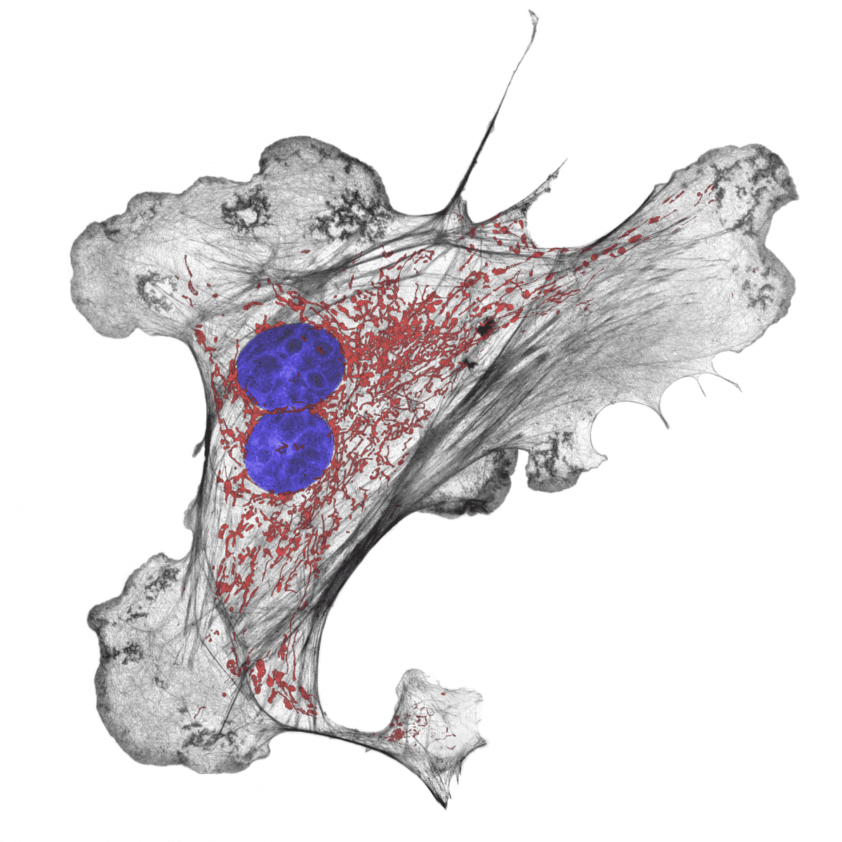
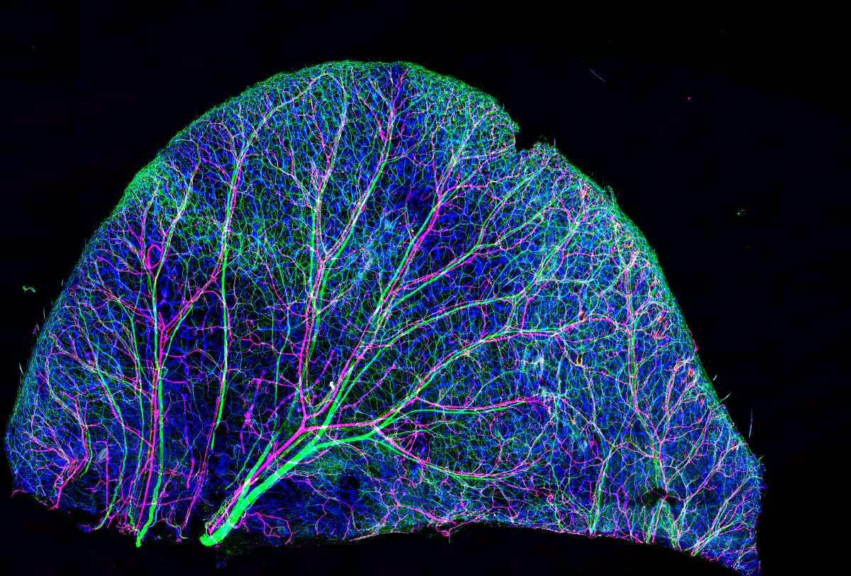
Lung artery cell
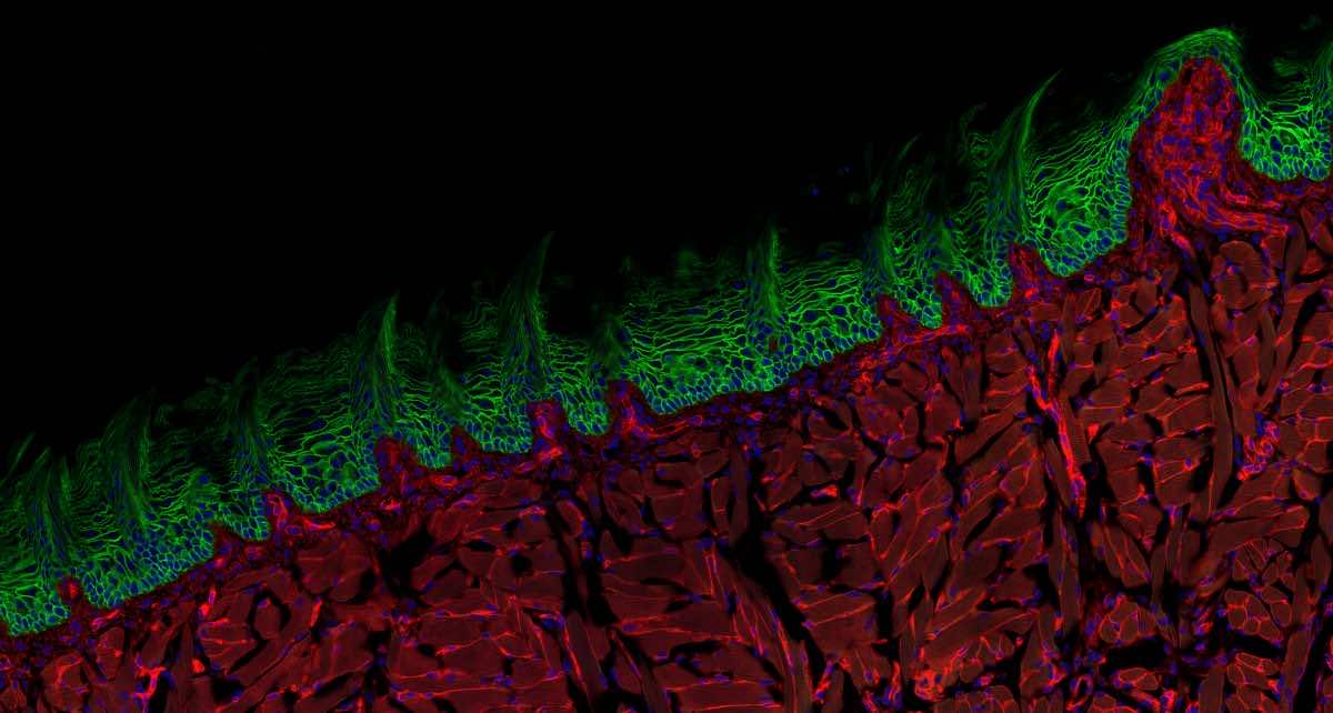
Rat tongue’s cross section
Closeup of ancient Chinese pottery
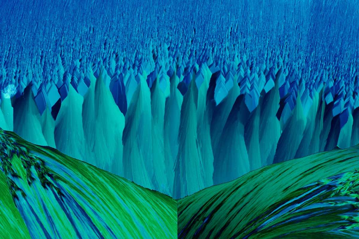
Plant producing crystals to defend itself

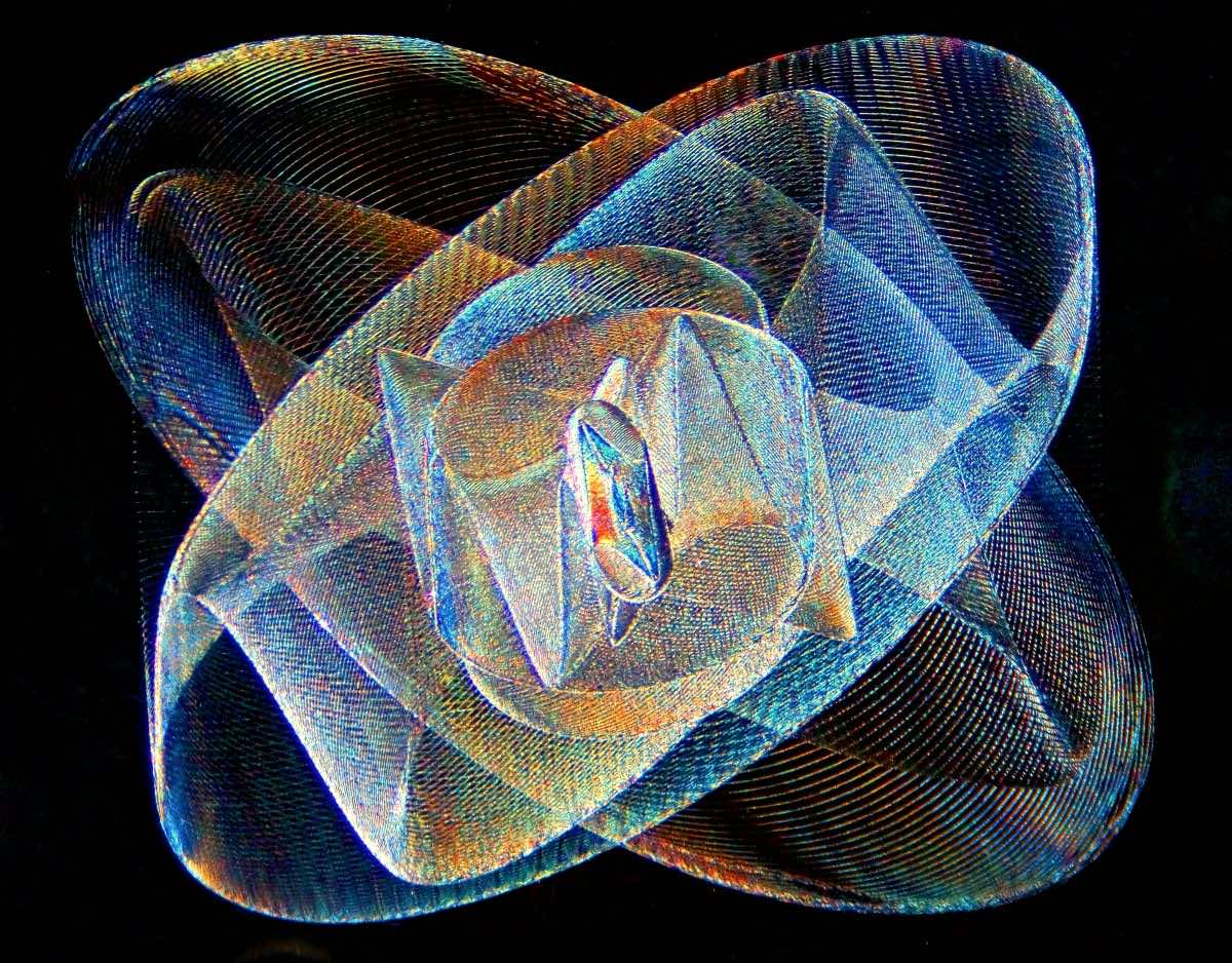
Engraving on a lens-based microscope from 1880
Cross section of fern
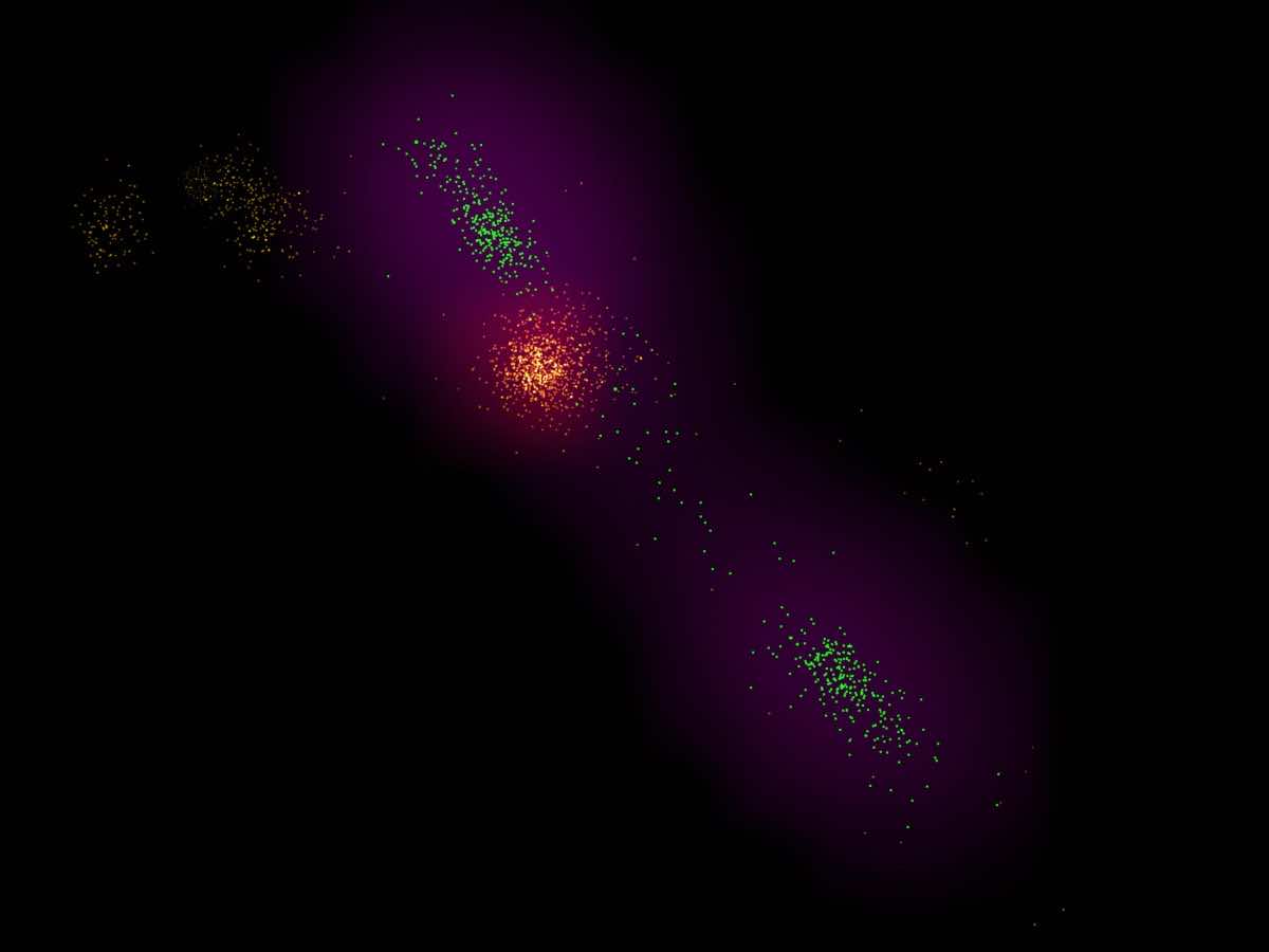
Bacterial DNA and probe
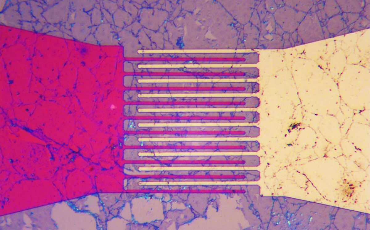
Gold and Titanium electrodes
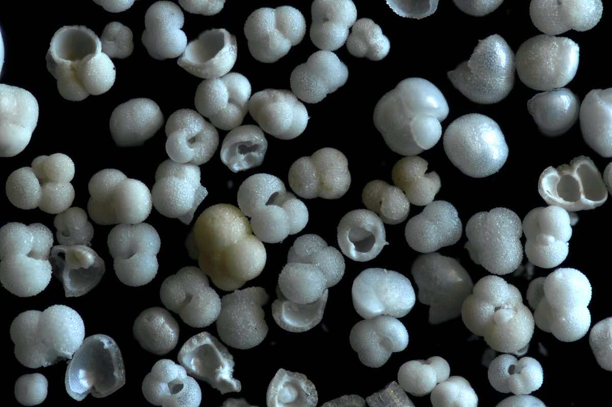
Shells from the sea
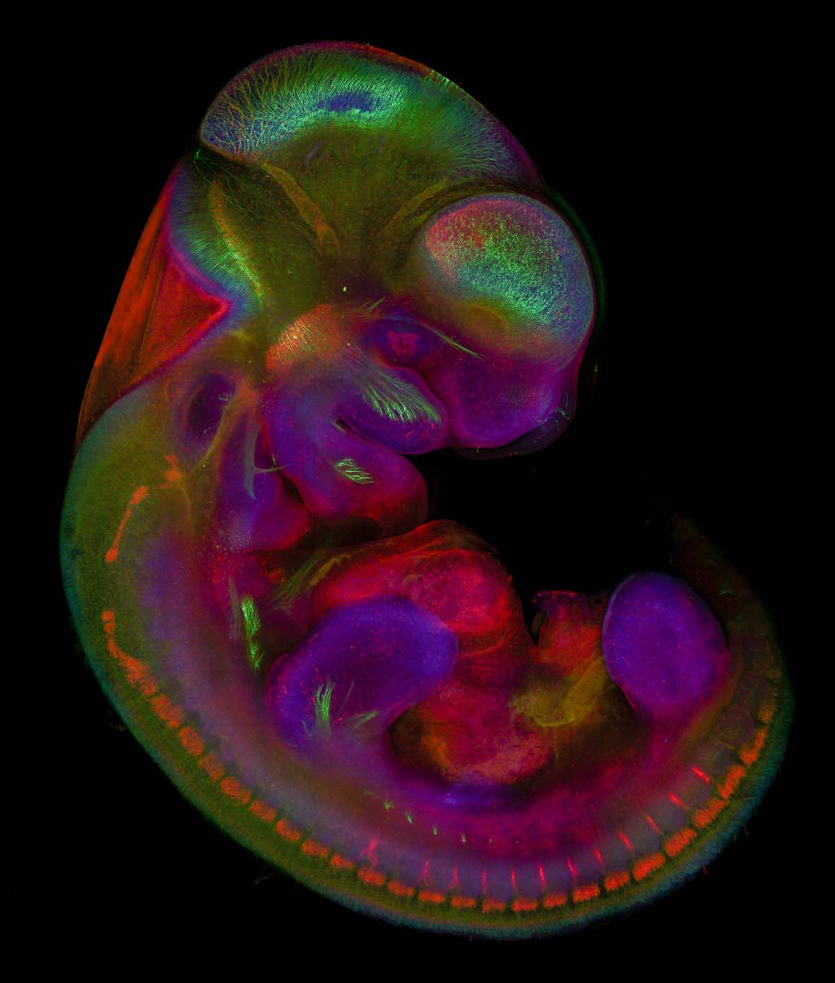
Muse embryo
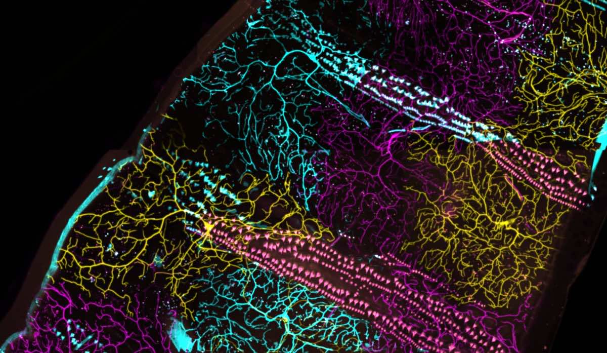
Colored neurons in fly larva
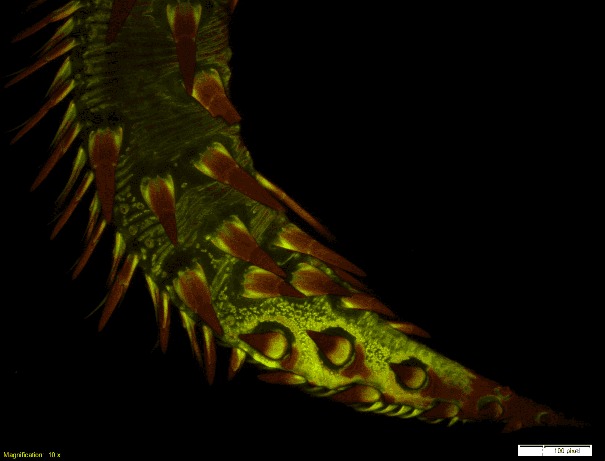
Beetle’s foreleg
Skin of a shelled animal
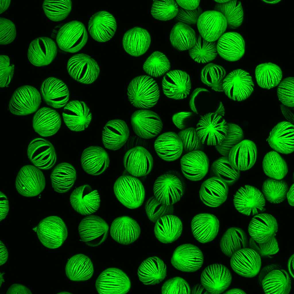
Pollens of a lily
Neural stem cells from human body
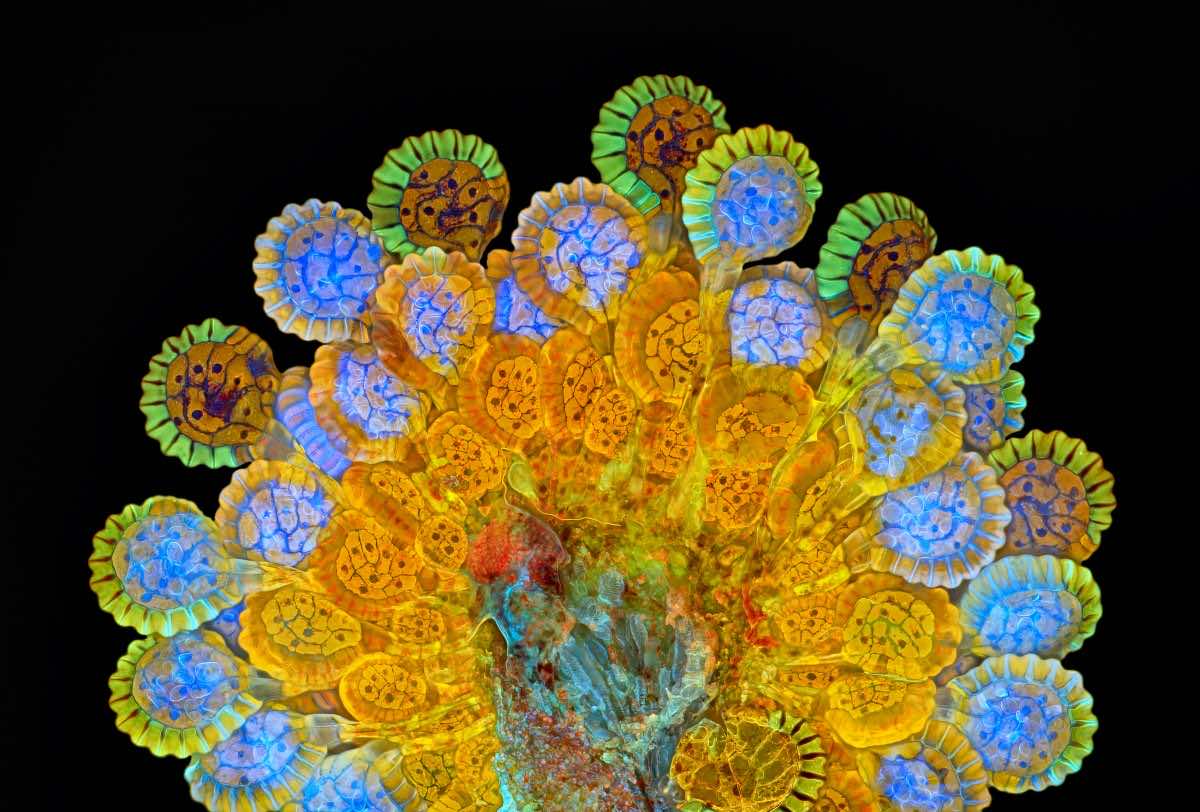
Fern Sorus at varying degrees of maturity

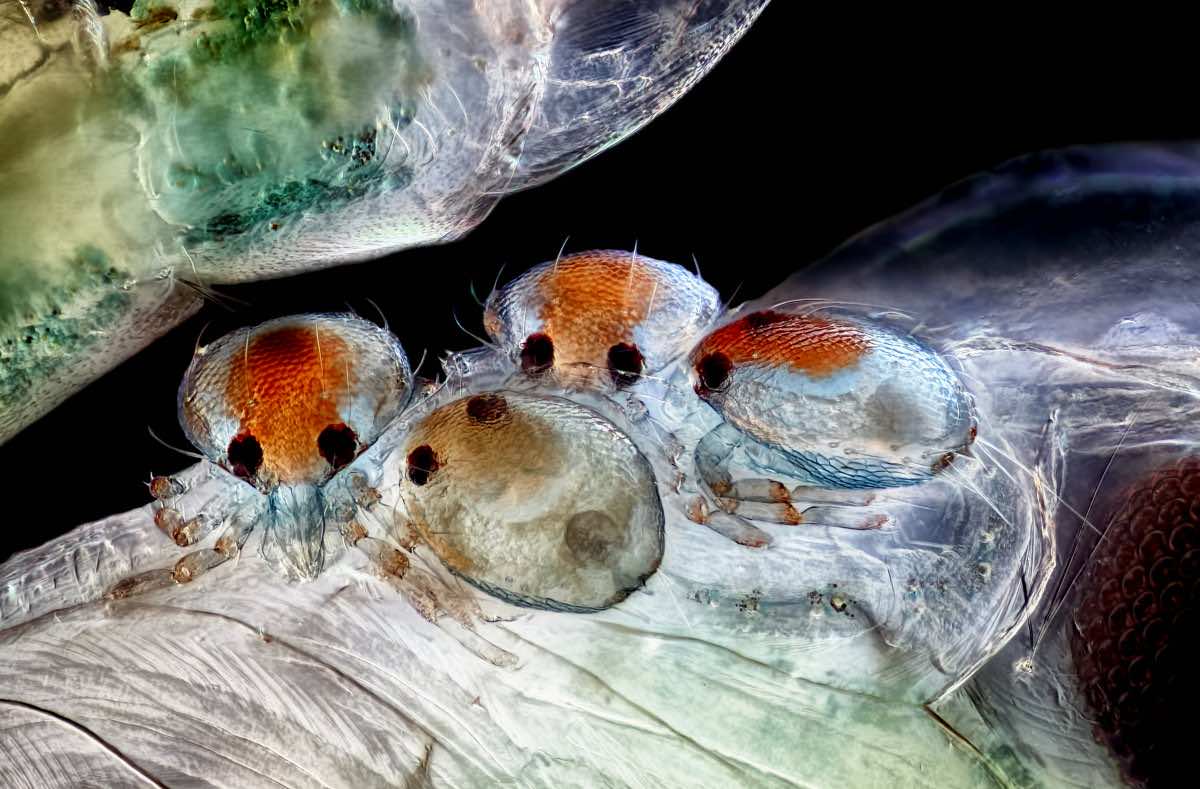
Mites on an insect pupae
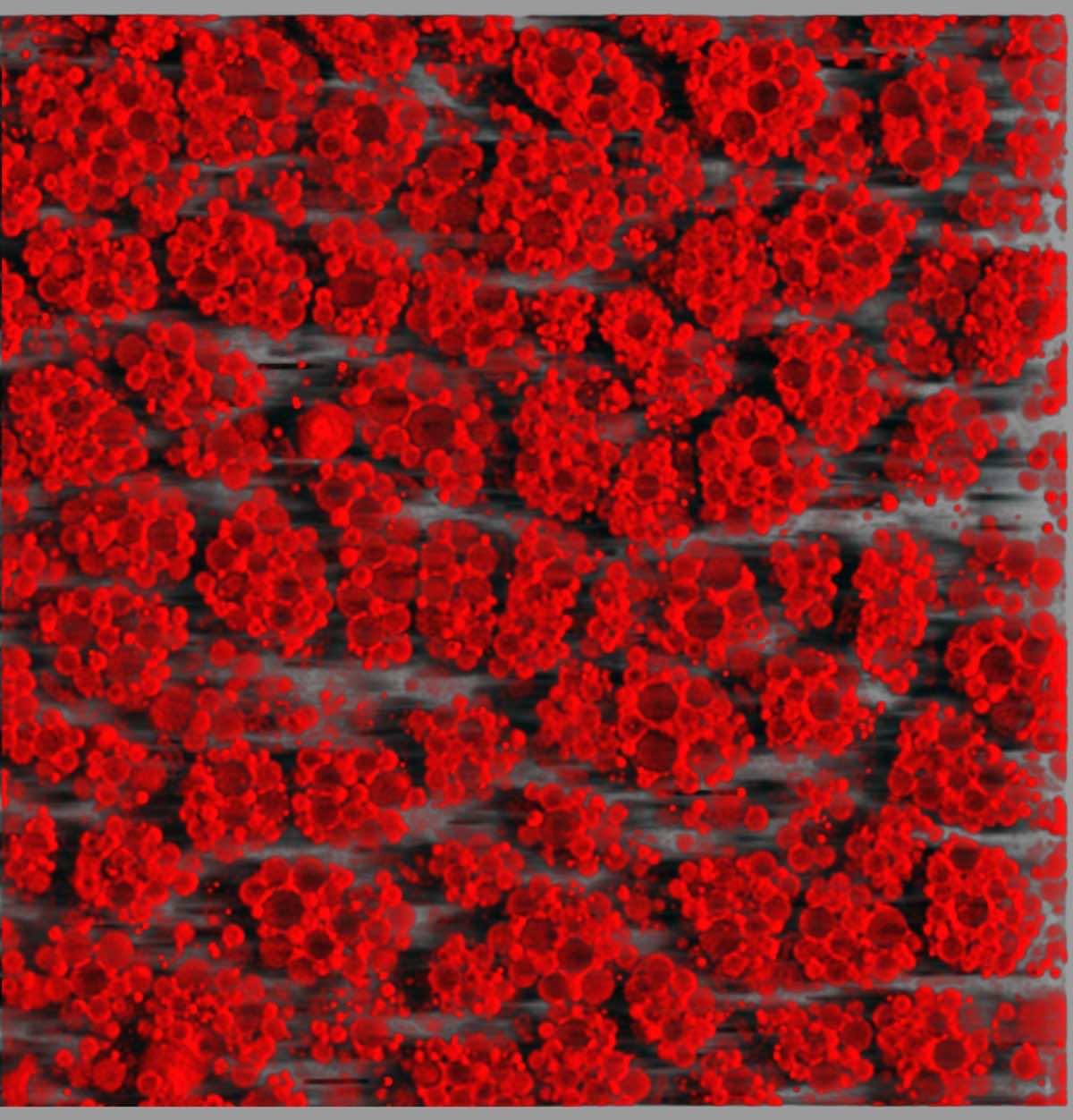
3-D rendering of mouse fat
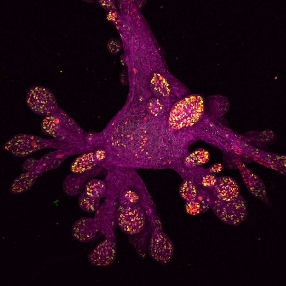
Bud of human breast gland
Shells from the pacific ocean
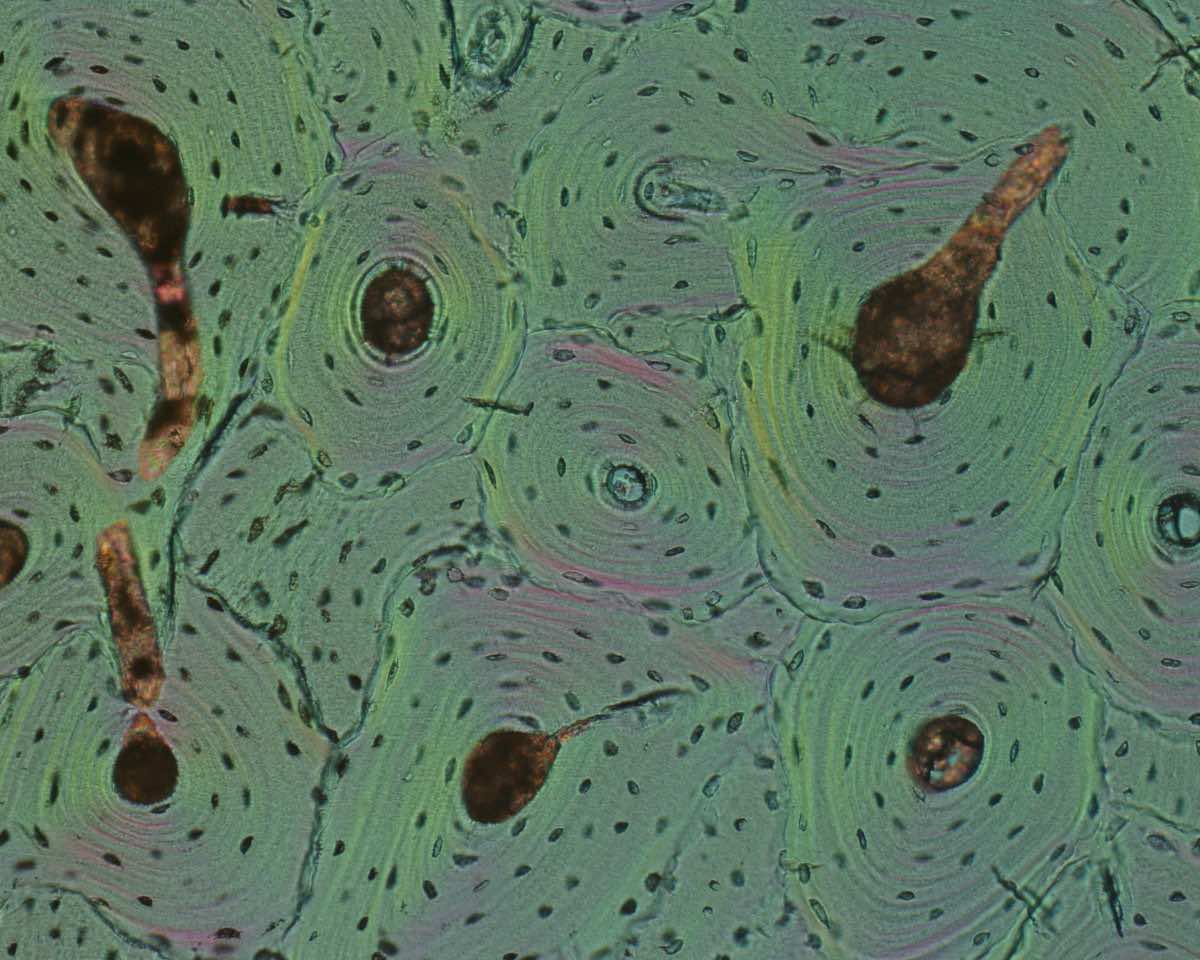
Nerves and blood vessels;s near the skin
